Athelia salicum Pers.
Basidiomycota: Agaricomycotina: Agaricomycetes: Atheliales: Atheliaceae: Athelia ![]()
![]()
Introduction
Athelia salicum is a drab crust. Even crust experts fail to feel any excitement for it. However, it is common, and species in the genus Athelia are ecologically important as saprotrophs, pathogens, and ectomycorrhizal fungi. Even if one overcomes their initial boredom, Athelia is difficult to work with because of their simplicity, both macroscopic and microscopic, and the taxonomic importance of the few characteristics that are used to separate species have not been tested with molecular phylogenetic studies.
These specimens have occassional leptocystidia and smaller spores than A. salicum, which could warrant a new species description.
Description
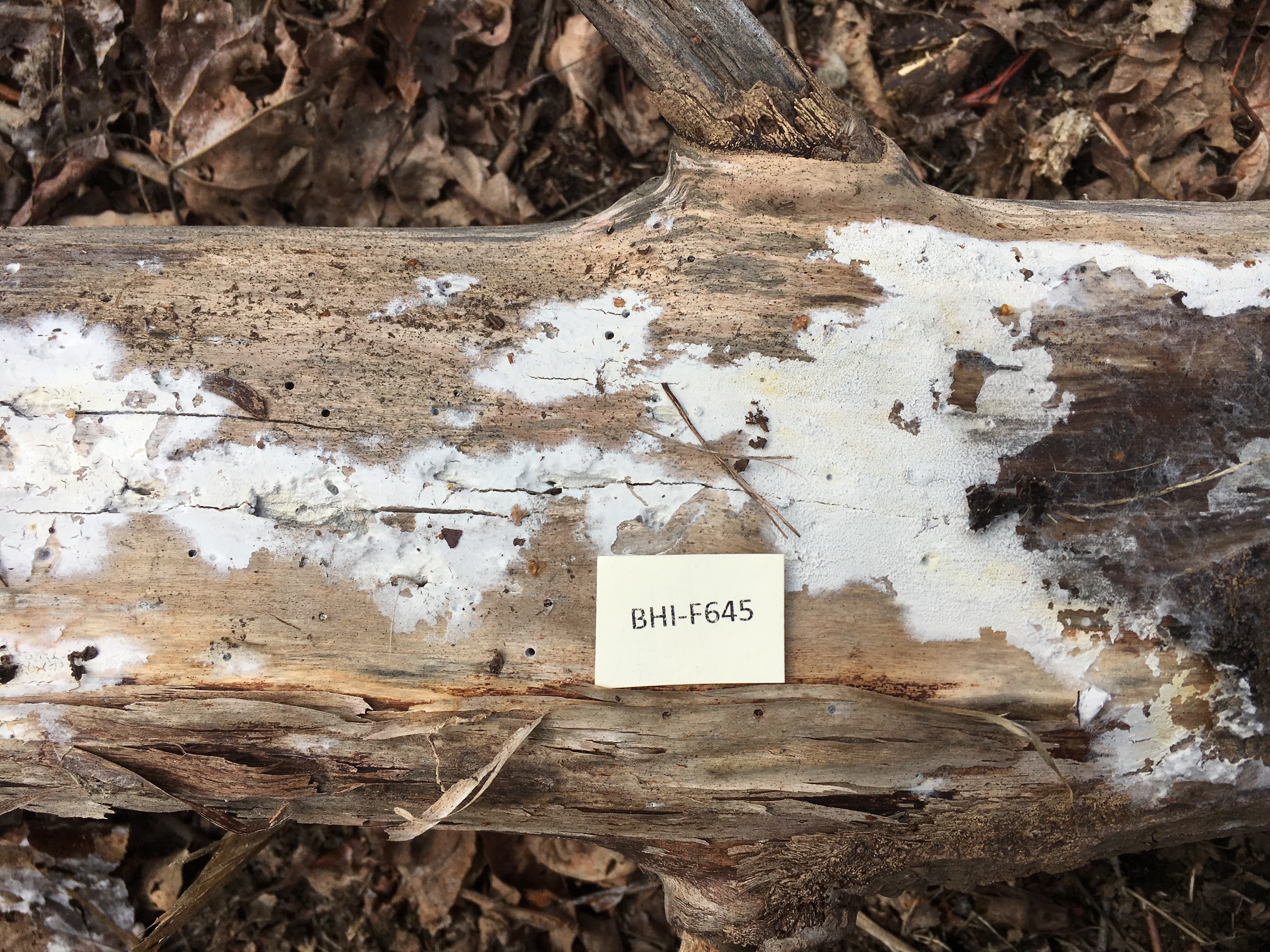
Ecology: The described specimens were found growing on standing and fallen hardwood trunks on two different islands at the Boston Harbor Islands National Recreation Area.
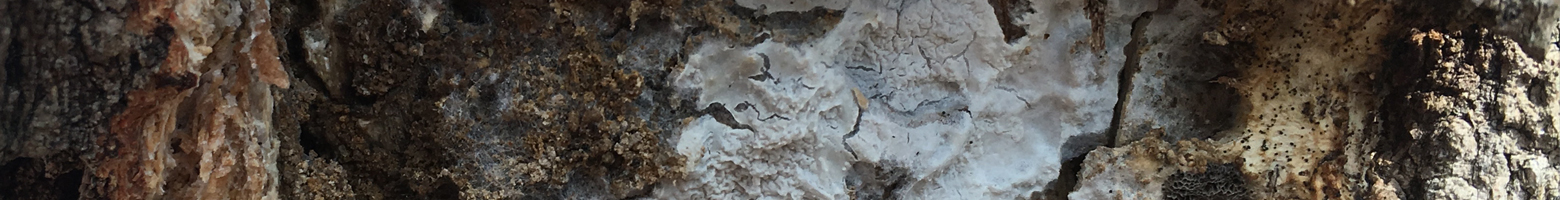
Basidiocarp: Effused, pellicular, easily peelable from the substrate; hymenial surface slightly merulioid when fresh, even when dry, whitish with yellow tints; margin arachnoid to byssoid.
Chemical reactions: NA
Spore print: NA
Hyphal system: Monomitic; subhymenial hyphae with simple septa, width (2.4) 3.0–4.1 (4.6) µm, x̄ = 3.6 µm (n = 10 per specimen); subicular hyphae with scattered clamps, slightly wider and with slightly thicker walls than the subhymenial hyphae, width (3.2) 3.8–5.0 (6.0) µm, x̄ = 4.4 µm (n = 10 per specimen), with acicular and blocky, rectangular crystals scattered on the subicular hyphae.
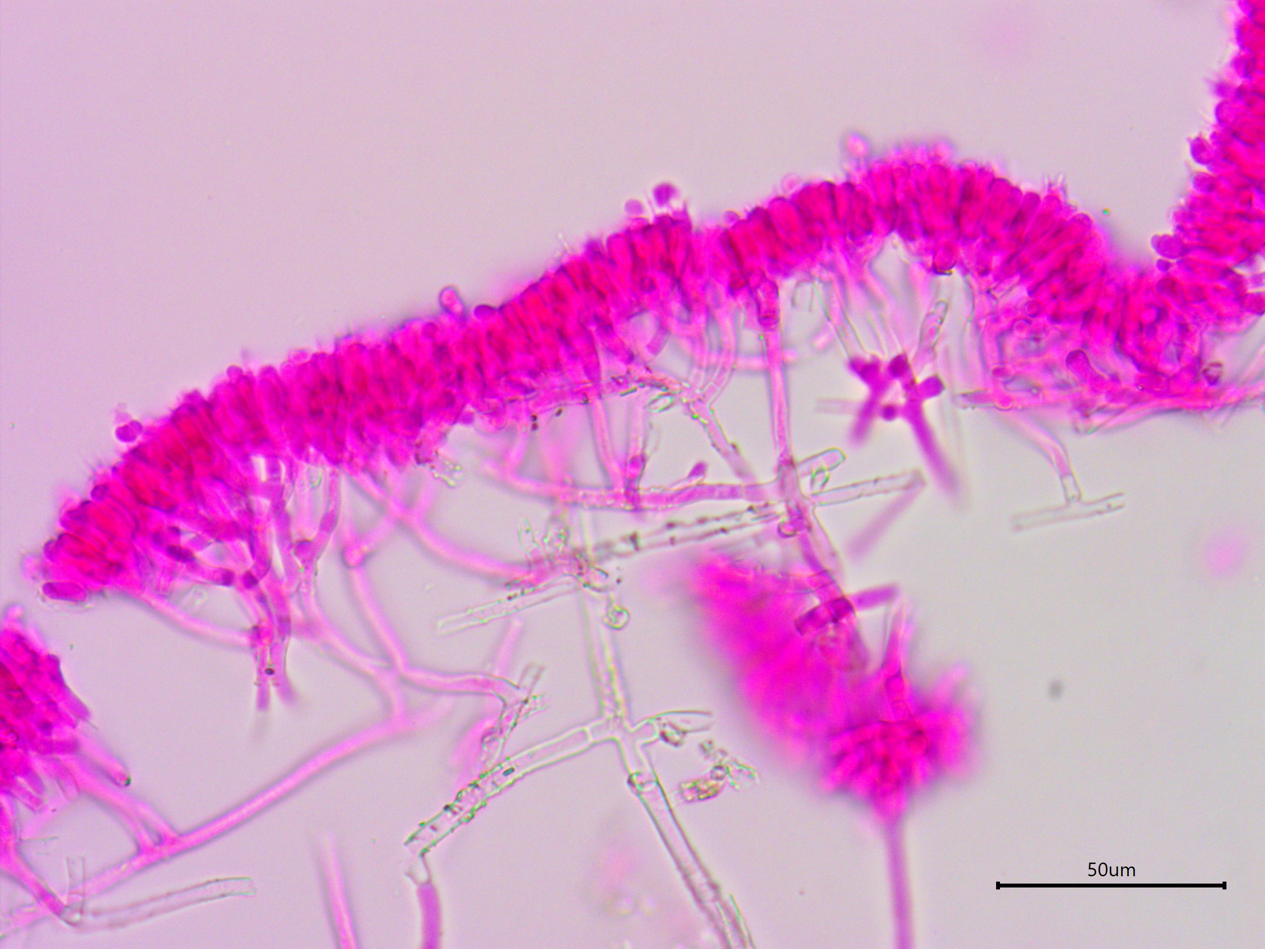
Basidia: Terminal, clavate, without a basal clamp; length (12.2) 13.7–18.4 (21.1) µm, width (4.3) 4.6–5.5 (5.9) µm, x̄ = 16.1 ✕ 5.0 µm (n = 10 per specimen), with four sterigmata, length (2.7) 3.2–4.7 (5.5) µm, x̄ = 3.9 µm (n = 10 per specimen).
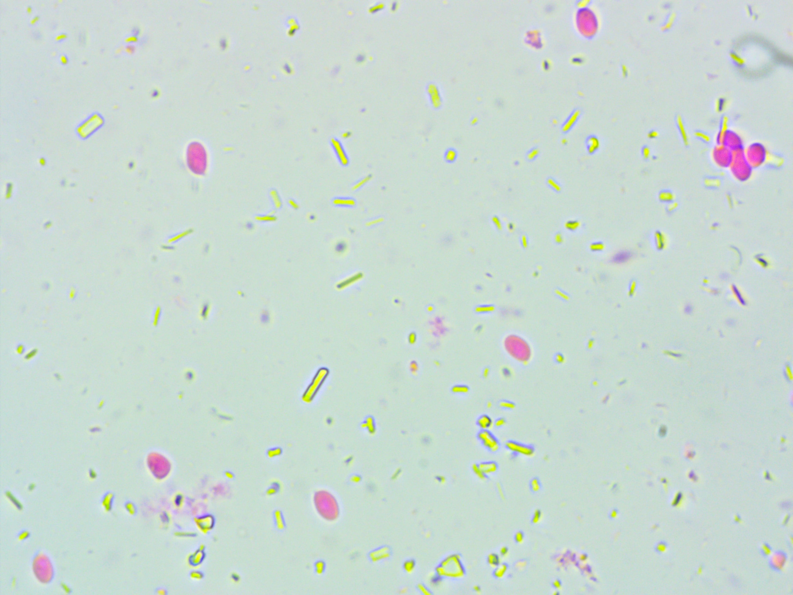
Basidiospores: Thin-walled, hyaline, inamyloid, acyanophilous, cylindrical; length (4.5) 5.2–6.2 (6.8) µm, width (2.4) 2.8–3.6 (4.2) µm, x̄ = 5.7 ✕ 3.2 µm, Q (1.4) 1.6–2.0 (2.4), sometimes agglutinated in pairs or in groups of four (n = 30 per specimen).
Sterile structures: Thin-walled leptocystidia or cystidioles infrequent, projecting up to 25 µm beyond the hymenial surface.
Sequences: ITS rDNA (MF161310, MF161314).
Notes: All measurements were taken using a smash mount of dried tissue in 5% KOH stained with phloxine. The presence of leptocystidia is a defining characteristic of Athelia cystidiolophora but the presence of clamps excludes that possibility.
Specimens Analyzed
BHI-F0636; 21 March 2017; Slate Island, Boston Harbor Islands National Recreation Area, MA, USA, 42.2678 -70.9125; leg. Alden Dirks & Lara Kappler, det. Alden Dirks, ref. Jülich & Stalpers (1980); Farlow Fungarium ——.
BHI-F0645, MO353240; 21 March 2017; Grape Island, Boston Harbor Islands National Recreation Area, MA, USA, 42.2682 -70.9225; leg. Alden Dirks & Lara Kappler, det. Alden Dirks, ref. Jülich & Stalpers (1980); Farlow Fungarium ——.
References
Jülich, W. & Stalpers, J. A. (1980). The Resupinate Non-poroid Aphyllophorales of the Temperate Northern Hemisphere. North-Holland Publishing Company, Amsterdam, Oxford, New York.
Links
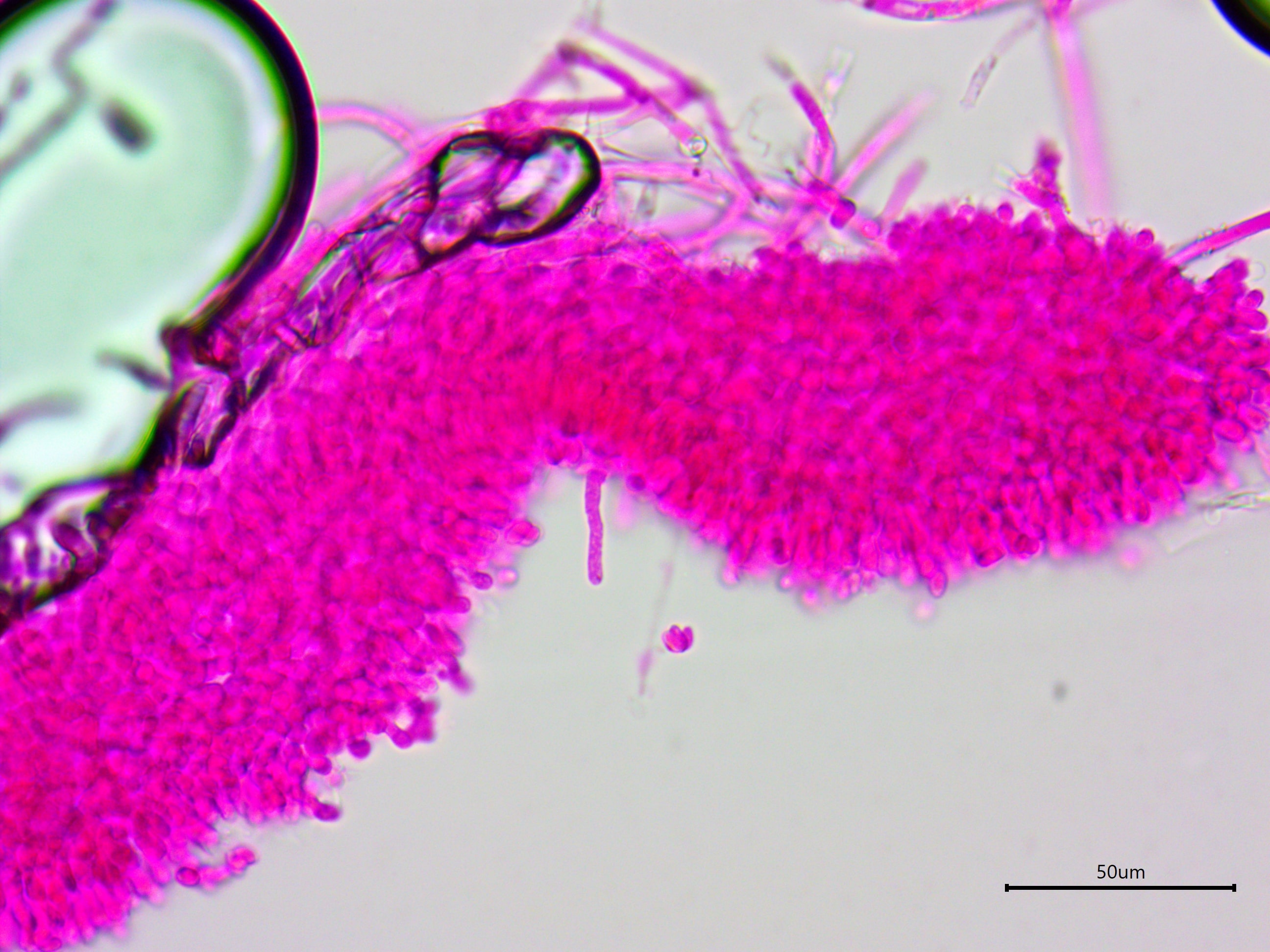
Athelia salicum is found on decaying wood and can spread across a large area. When fresh, it has a merulioid texture.
The fruiting body is very thin, consisting of a hymenium and a loose subicular network.
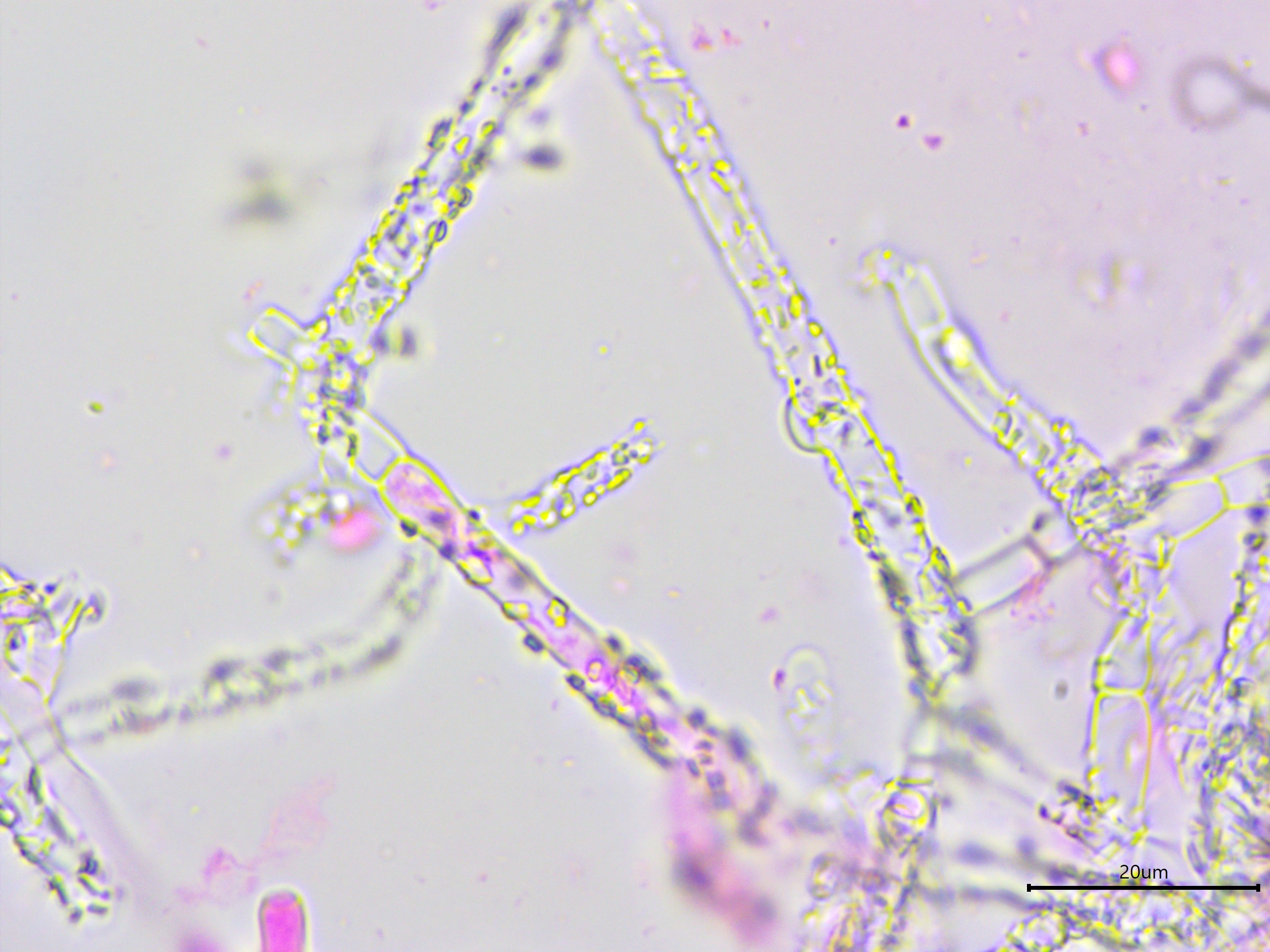
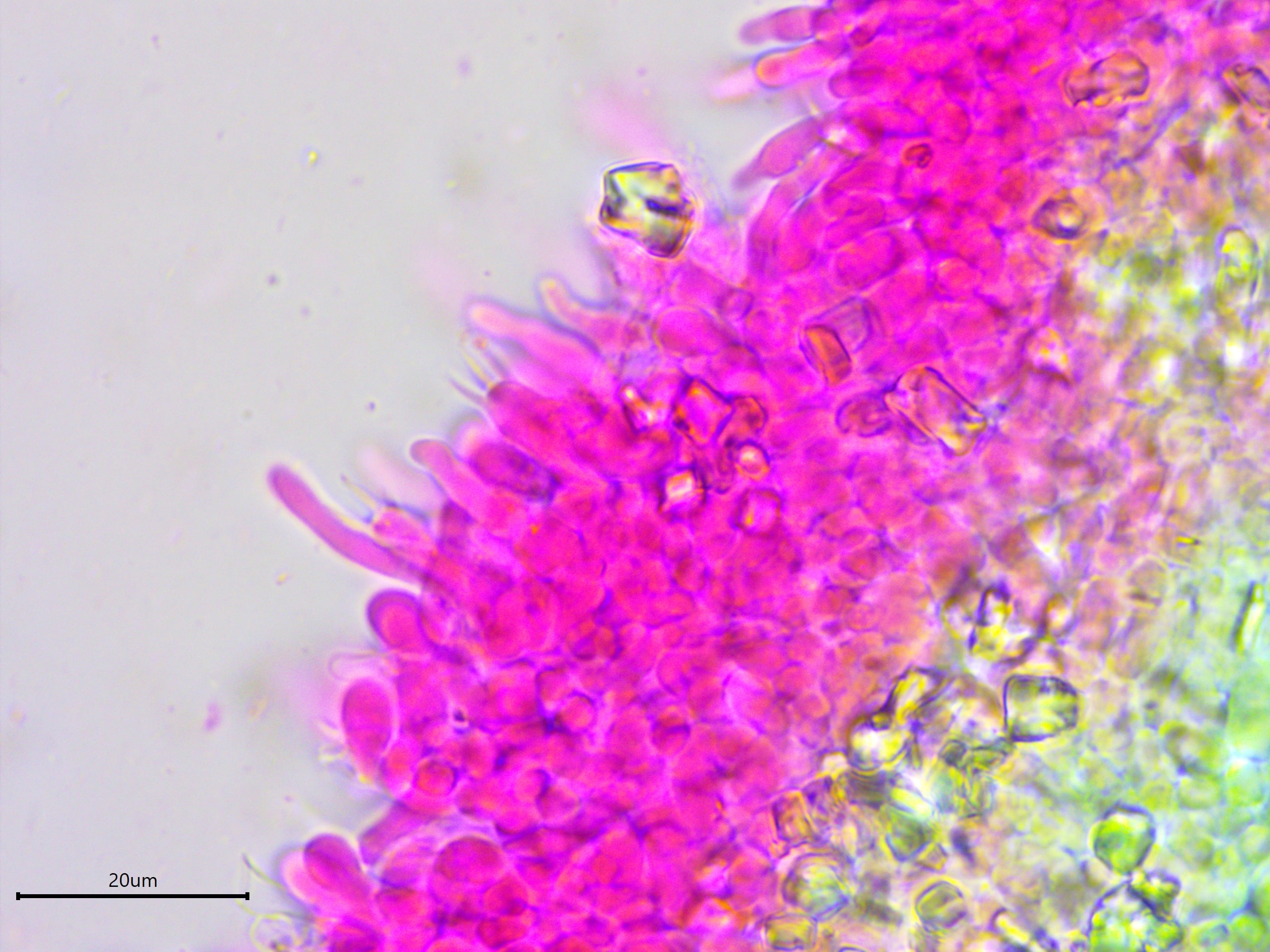
The subicular hyphae are wider and thicker-walled than the subhymenial hyphae, have occassional clamp connections, and are encrusted.
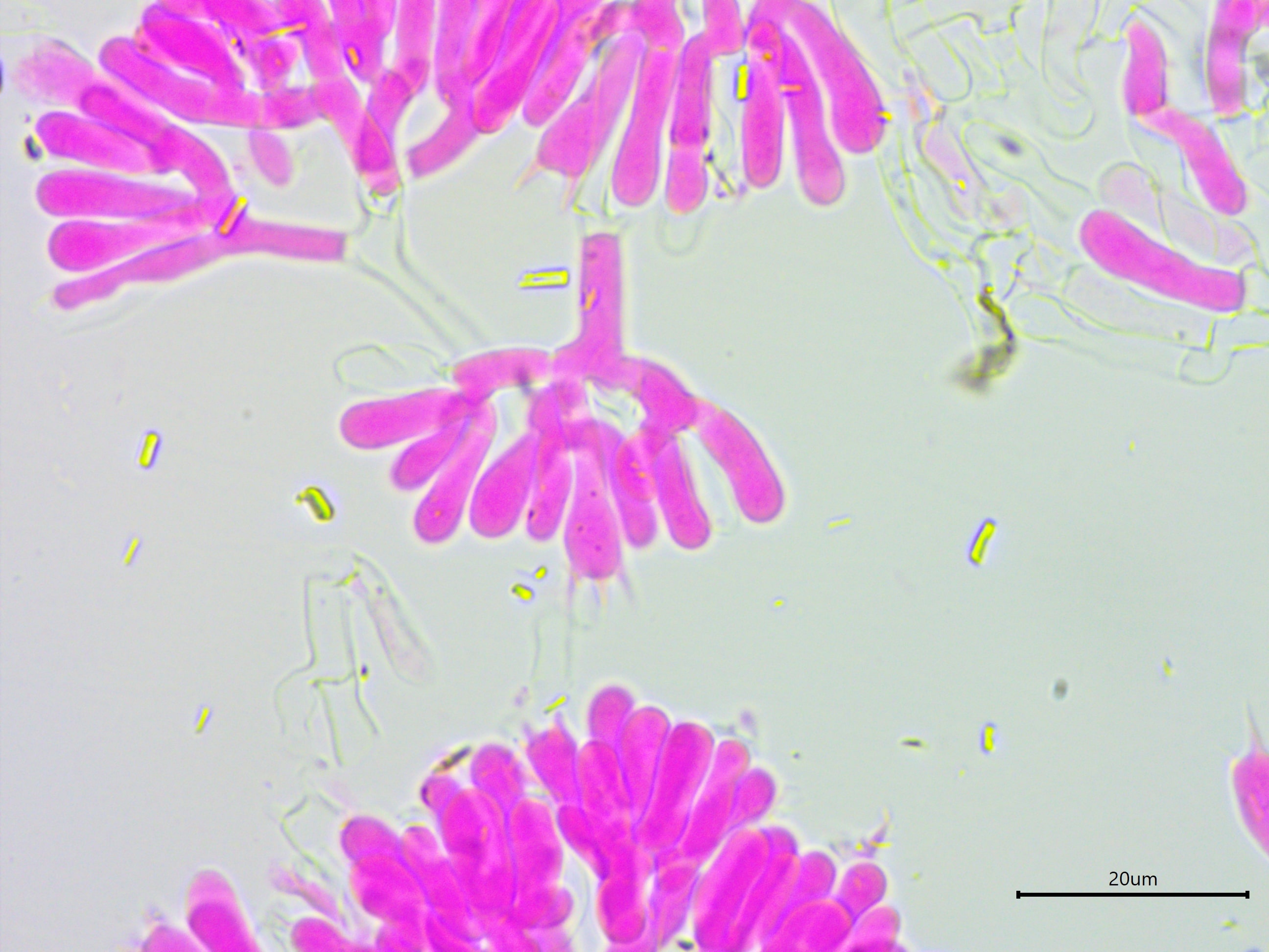
The basidia are in a candelabra arrangement, typical for Athelia spp.
Athelia salicum has cylindrical spores with a distinct apiculus.
Occassionally, leptocystidia were seen projecting beyond the hymenium.
This is another look at a projecting leptocystidium.

Here is another look at Athelia salicum leptocystidia.
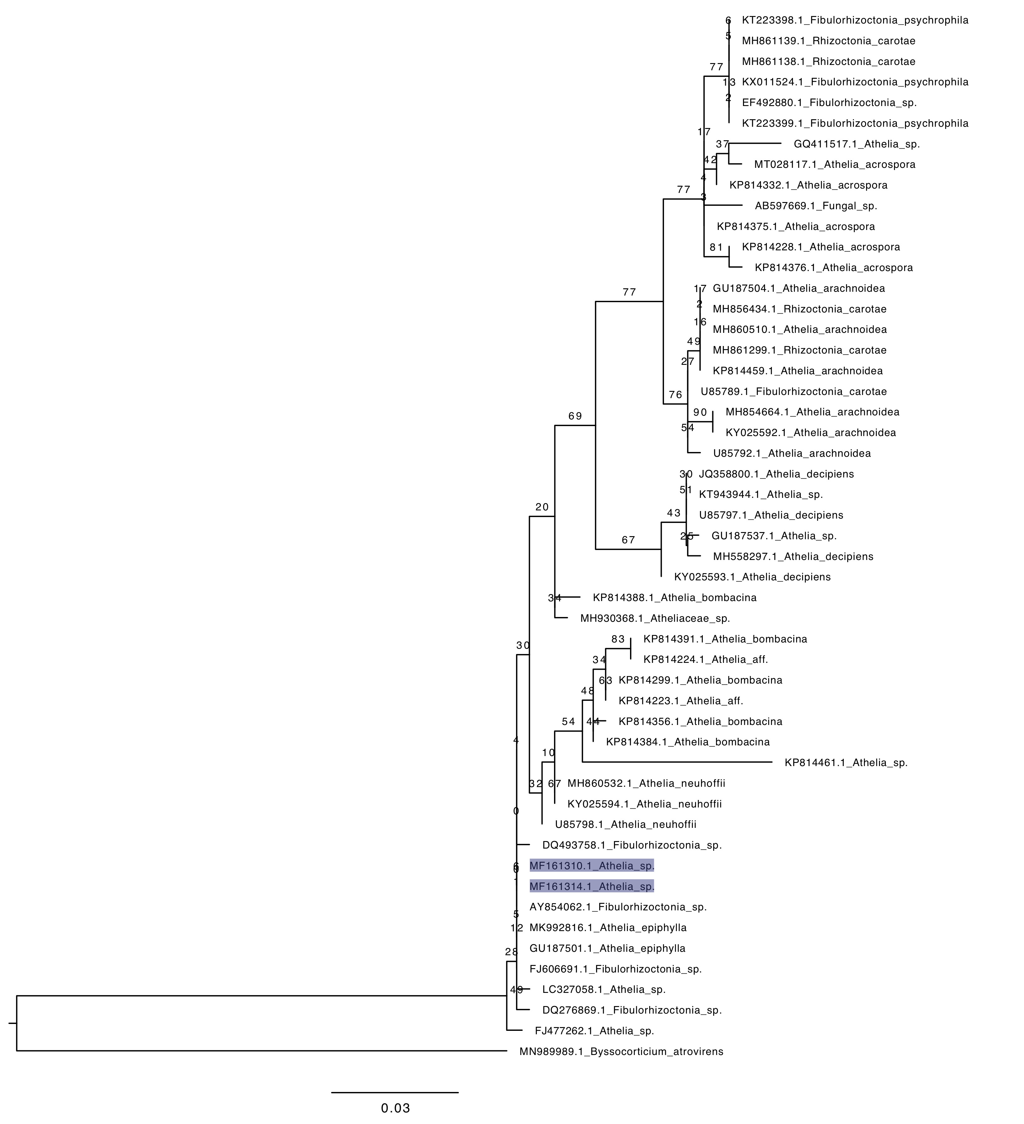
Phylogenetic tree of ITS rDNA sequences from the studied specimen (highlighted) and selected vouchered specimens on GenBank. The sequences were processed with ITSx to remove the flanking SSU and LSU partial sequences, aligned in SeaView with MUSCLE, cleaned up with GBlocks, and made into a tree with RAxML using 100 bootsrap replicates and the GTRGAMMA substitution model. Byssocorticium atrovirens serves as the outgroup. Note that ITS does not do a good job at resolving the phylogeny of this clade.