Ceriporia spissa (Schwein. ex Fr.) Rajchenb.
Basidiomycota: Agaricomycotina: Agaricomycetes: Polyporales: Irpicaceae: Ceriporia ![]()
![]()
Introduction
Ceriporia species are resupinate, annual polypores that produce a white rot and are typically brightly colored. About 80 species are distributed worldwide (Chen et al. 2020). Ceriporia spissa as defined morphologically is relatively easy to recognize by its orange color, resupinate growth, and small pores. However, phylogenetic analyses show that the morphological species concept of Ceriporia spissa is polyphyletic so further taxonomic research is definitely needed (Jia et al. 2014). Fortunately, the type specimen is from Massachusetts, so northeastern collections have the best chance of being the "true" Ceriporia spissa.
A fair number of crusts or crust-like species share the orangish coloration of Ceriporia spissa and are commonly misidentified as such on iNaturalist (probably due to their auto-identification algorithm) but can be easily distinguished with a human brain by the lack of pores. Phanerochaete chrysorhiza produces extensive rhizomorphs and has an odontioid to hydnoid hymenophore. Phlebia coccineofulva is typically a brighter red color when fresh and has a merulioid to grandinoid hymenophore. Stereum complicatum is an orange effused-reflexed parchment fungus. Pyronema omphalodes is a bright orange crustose ascomycete that grows on recently burned ground. Another potential lookalike is not a single species at all but rather a consortium of microbes. In the spring, sap gets flowing and can ooze out of cuts in tree trunks or the stumps of recently felled trees, upon which a slimy orange mass may form. This slime is composed of yeasts, filamentous fungi, and bacteria that are said to feed on the sugary sap. The orange color is primarily a result of carotene production by the yeast Cryptococcus macerans.
Description
Ecology: Ceriporia spissa grows as a saprotroph on the underside of hardwood logs in northeastern North America, but is also reported from western United States, tropical South America (Coelho et al. 2005), and China (Jia et al. 2014).
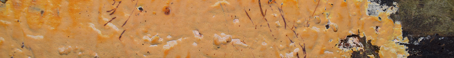
Basidiocarp: Resupinate, effused, poroid fruiting body extending over a meter on the side of a large fallen hardwood trunk; egg-yolk yellow, salmon orange, or bright orange, bruising vinaceous red, drying darker brownish orange, sometimes with a thin white margin but mostly without a differentially colored margin; hymenophore ceraceous with 5-7 pores per mm; hymenium subtended by a vinaceous context.
Chemical reactions: NA
Spore print: White.
Hyphal system: Monomitic, all hyphae with simple septa; subicular hyphae thick-walled; subhymenial hyphae thin-walled tightly running parallel and difficult to discern, coated with small granules; subicular hyphae width (2.8) 2.8–3.6 (3.9) µm, x̄ = 3.2 µm; subicular hyphae wall thickness (0.8) 0.6–1.3 (2) µm, x̄ = 1 µm; subhymenial hyphae width (2.8) 3–3.7 (3.8) µm, x̄ = 3.3 µm; 10 subicular hyphae and 10 subhymenial hyphae measured in KOH stained with phloxine.
Basidia: Clavate with four sterigmata and no basal clamp; basidia length (9.6) 10.5–15.5 (18) µm, width (3.8) 4–4.8 (5) µm, x̄ = 13 ✕ 4.4 µm, Q (2) 2.3–3.7 (4.7), x̄ = 3; sterigmata length (2.2) 2.3–3.1 (3.3) µm, x̄ = 2.7 µm; 10 basidia and sterigmata measured in KOH, with or without phloxine.
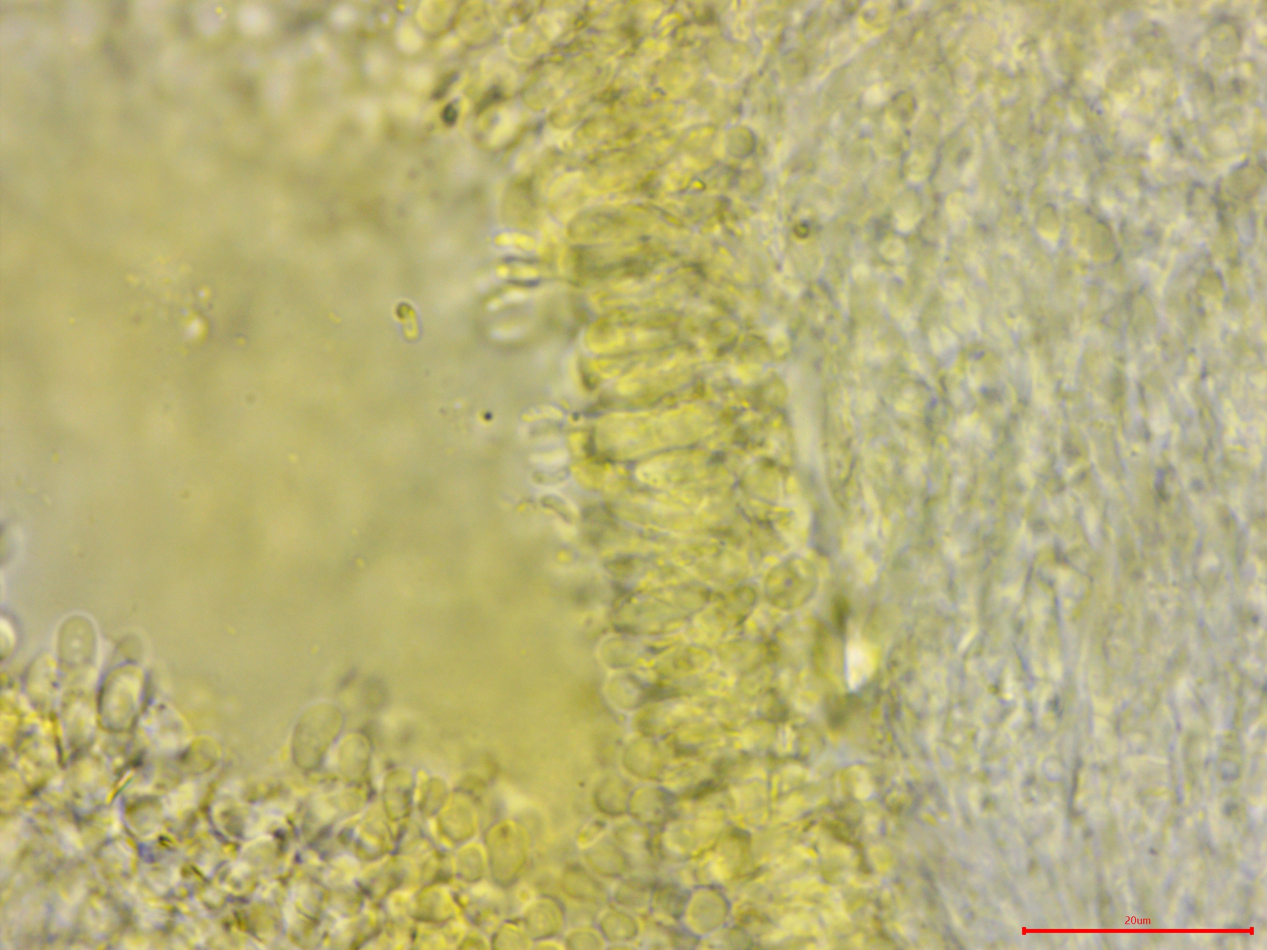
Basidiospores: Narrowly cylindrical, straight to curved, small, hyaline, inamyloid; length (3.7) 4.2–4.9 (5) µm, width (1.3) 1.5–1.8 (2) µm, x̄ = 4.6 ✕ 1.6 µm, Q (2.4) 2.6–3.1 (3.4), x̄ = 2.8; 30 basidiospores measured in tap water.
Sterile structures: Absent.
Sequences: ITS rDNA (MZ919231).
Specimens Analyzed
ACD0304, MO413092; 11 June 2020; Eberwhite Nature Area, Washtenaw Co., MI, USA, 42.2725 -83.7678; leg. Alden Dirks, det. Alden Dirks, recognized by sight and ref. Spirin et al. (2016); University of Michigan Fungarium MICH 352265.
References
Chen, C.-C., Chen, C.-Y., Lim, Y. W., & Wu, S.-H. (2020). Phylogeny and taxonomy of Ceriporia and other related taxa and description of three new species. Mycologia, 112(1), 64–82.
Coelho, G., Reck, M., Borges da Silveira, R. M., & Trinidad Guerrero, R. (2005). Ceriporia spissa (Schwein. ex Fr.) Rajchenb. (Basidiomycota): First record from Brazil. Biociências, 13(2), 107–111.
Jia, B.-S., Zhou, L.-W., Cui, B.-K., Rivoire, B., & Dai, Y.-C. (2014). Taxonomy and phylogeny of Ceriporia (Polyporales, Basidiomycota) with an emphasis of Chinese collections. Mycological Progress, 13(1), 81–93.
Spirin, V., Vlasák, J., Rivoire, B., Kout, J., Kotiranta, H., & Miettinen, O. (2016). Studies in the Ceriporia purpurea group (Polyporales, Basidiomycota), with notes on similar Ceriporia species. Cryptogamie, Mycologie, 37(4), 421–435.
Links
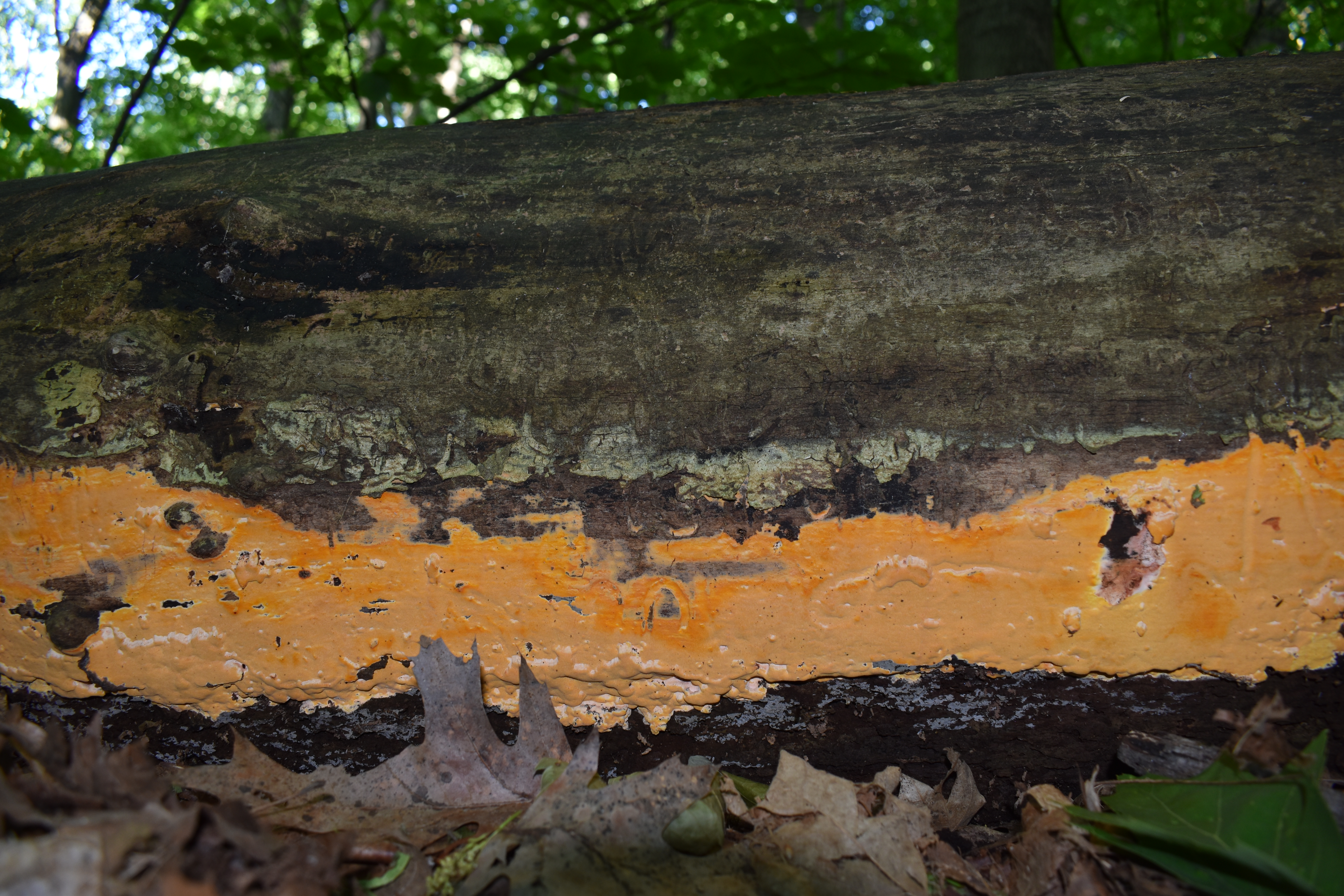
Extensive fruiting of Ceriporia spissa on the bottom edge and underside of a hardwood log.


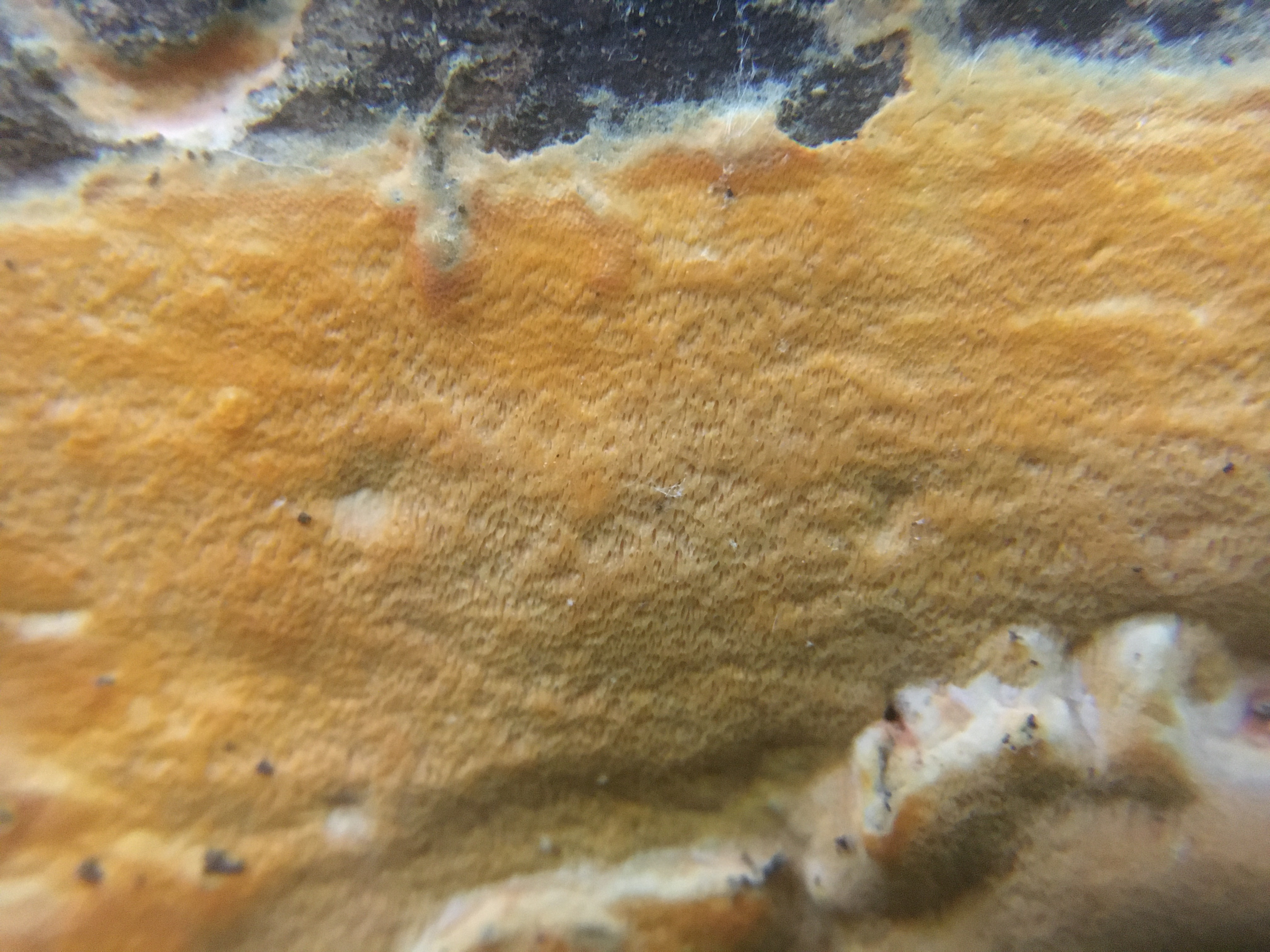
Ceriporia spissa has small pores, approx. 5-7 per mm.

The subhymenial tissue is vinaceous in color.
The spores are small and slightly curved.

Another look at a club-shaped to cylindrical basidium.
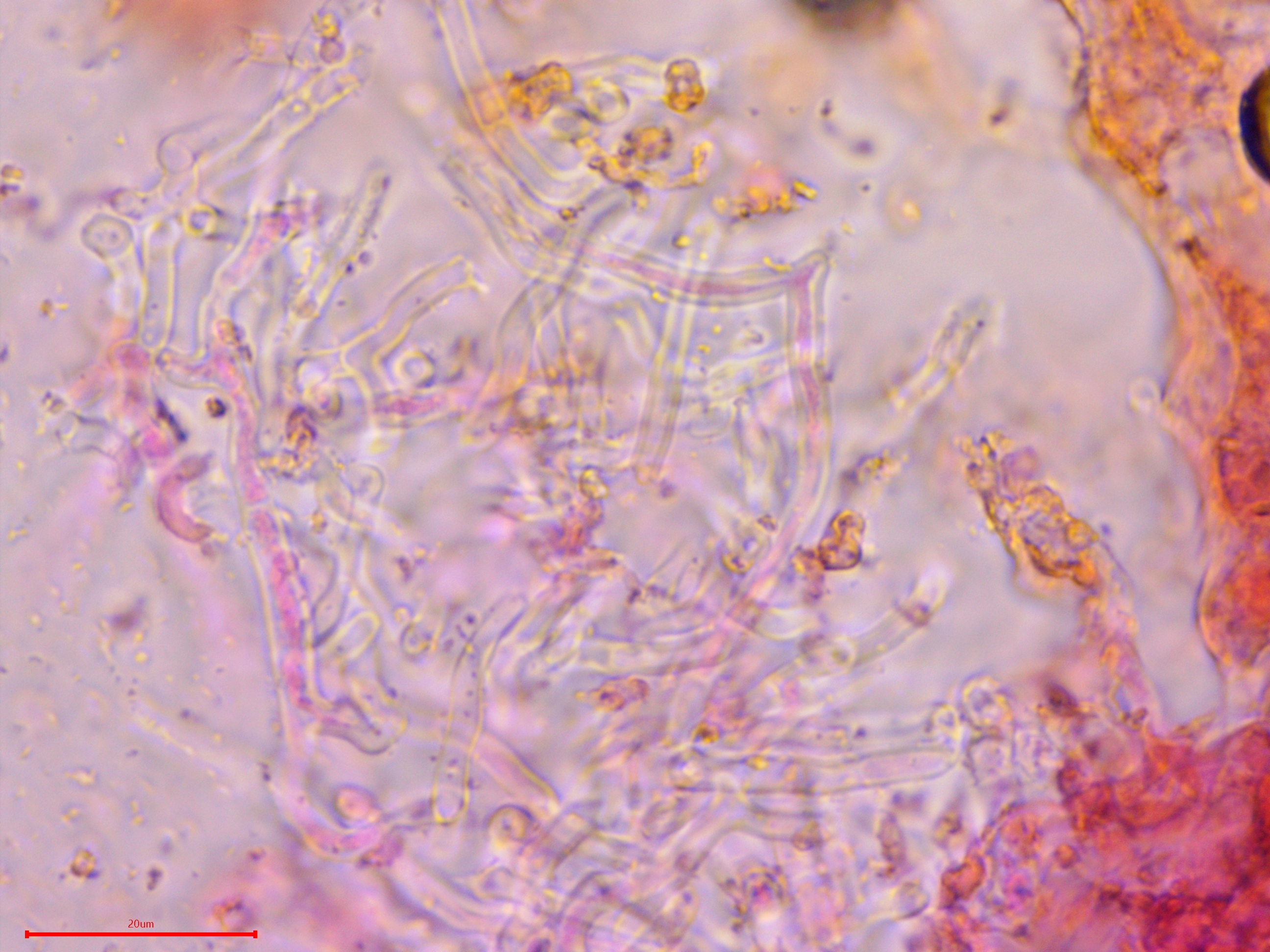
The subicular hyphae were notably thick walled. This image might show one clamp, but generally speaking there were none.
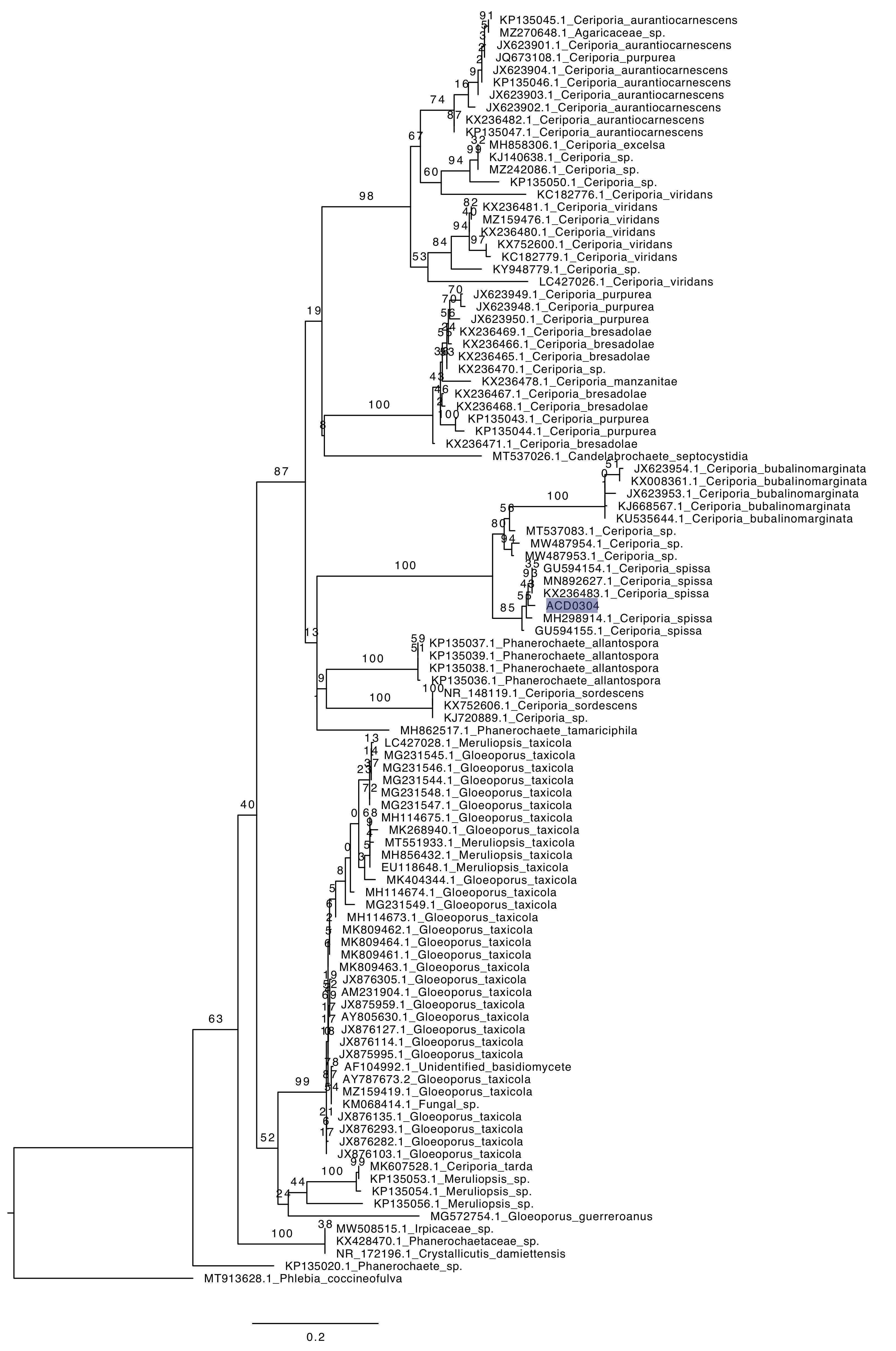
Phylogenetic tree of ITS rDNA sequences from the studied specimen (highlighted) and selected vouchered specimens on GenBank. The sequences were processed with ITSx to remove the flanking SSU and LSU partial sequences, aligned in SeaView with MUSCLE, and made into a tree with RAxML using 100 bootsrap replicates and the GTRGAMMA substitution model. Phlebia coccineofulva serves as the outgroup.