Mycoacia uda (Fr.) Donk
Pale-Yellow Splash Tooth
Basidiomycota: Agaricomycotina: Agaricomycetes: Polyporales: Meruliaceae: Mycoacia ![]()
![]()
Introduction
When fresh, Mycoacia uda can be identified in the field by its waxy consistency (a handlens helps); its pale yellow, toothed hymenium; and the bright purple (perhaps red to dark brown depending on the base color of that particular specimen) reaction to potassium hydroxide. Under the microscope, the yellow resinous content (visible in water, dissolving in KOH) and basidiospore size will clinch your ID. It's a typical phlebioid fungus, meaning it starts out waxy but dries horny hard, has small inamyloid basidiospores, and a hymenium arranged in a dense palisade.
Phlebia and related fungi have received a lot of attention from the corticiologist Karen Nakasone. To give you a sense of the magnitude of the job (and hopefully a greater appreciation for the hard work of taxonomists), she analyzed almost 500 specimens in just one study on Mycoacia uda and five other phlebioid crusts (Nakasone 1997)! And that's just one of many dozens of publications that she has published over her career. I'd be very proud of myself if I looked at that number of specimens in a decade.
Mycoacia as a genus name is widely in use for toothed phlebioid crusts although experts consider it a synonym of Phlebia (Nakasone 1997, Gorjón 2020). Phlebia uda and Sarcodontia uda are synonyms.
Description
Ecology: Saprotrophic on hardwood branches causing a white rot; common and widespread.
Basidiocarp: Effused, odontioid to hydnoid; when fresh, ceraceous in texture and (cooked) egg-yolk yellow, yellowish, or ochraceous in color, hardening and fading tannish brown when dry; aculei conical, up to 1 mm long (up to 3 mm in the literature), about 3 per mm, with sharp or frayed-looking (denticulate) tips, composed of a central core of encrusted tramal hyphae that project through the hymenium at the tips of the aculei giving them a paler color, arising singularly or connected at the base with others; margin abrupt or somewhat fimbriate.
Chemical reactions: Reacting purple to ammonia and potassium hydroxide, not absorbing iron salts (negative reaction).
Spore print: White.
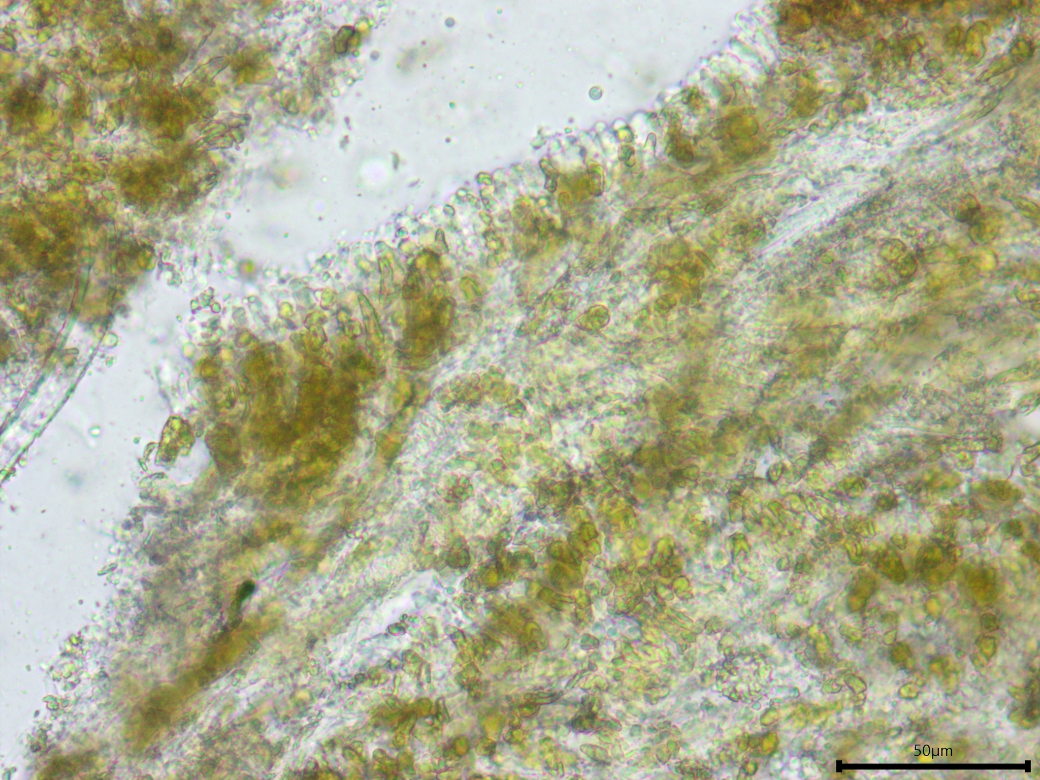
Hyphal system: Monomitic, apparently with clamp connections at all septa; tramal hyphae hyaline, thick-walled, densely encrusted with fine, acicular crystalline material, 2–3 µm wide.
Basidia: Cylindrical to narrowly clavate, terminal, with four sterigmata.
Basidiospores: Cylindrical to narrowly cylindrical, smooth, thin-walled, hyaline, inamyloid, sometimes guttulate; length (4.3) 4.8–5.5 (6.3) µm, width (2.2) 2.5–2.9 (2.9) µm, x̄ = 5.1 ✕ 2.7 µm, Q (1.6) 1.8-2.1 (2.3), x̄ = 1.9 (n = 30).
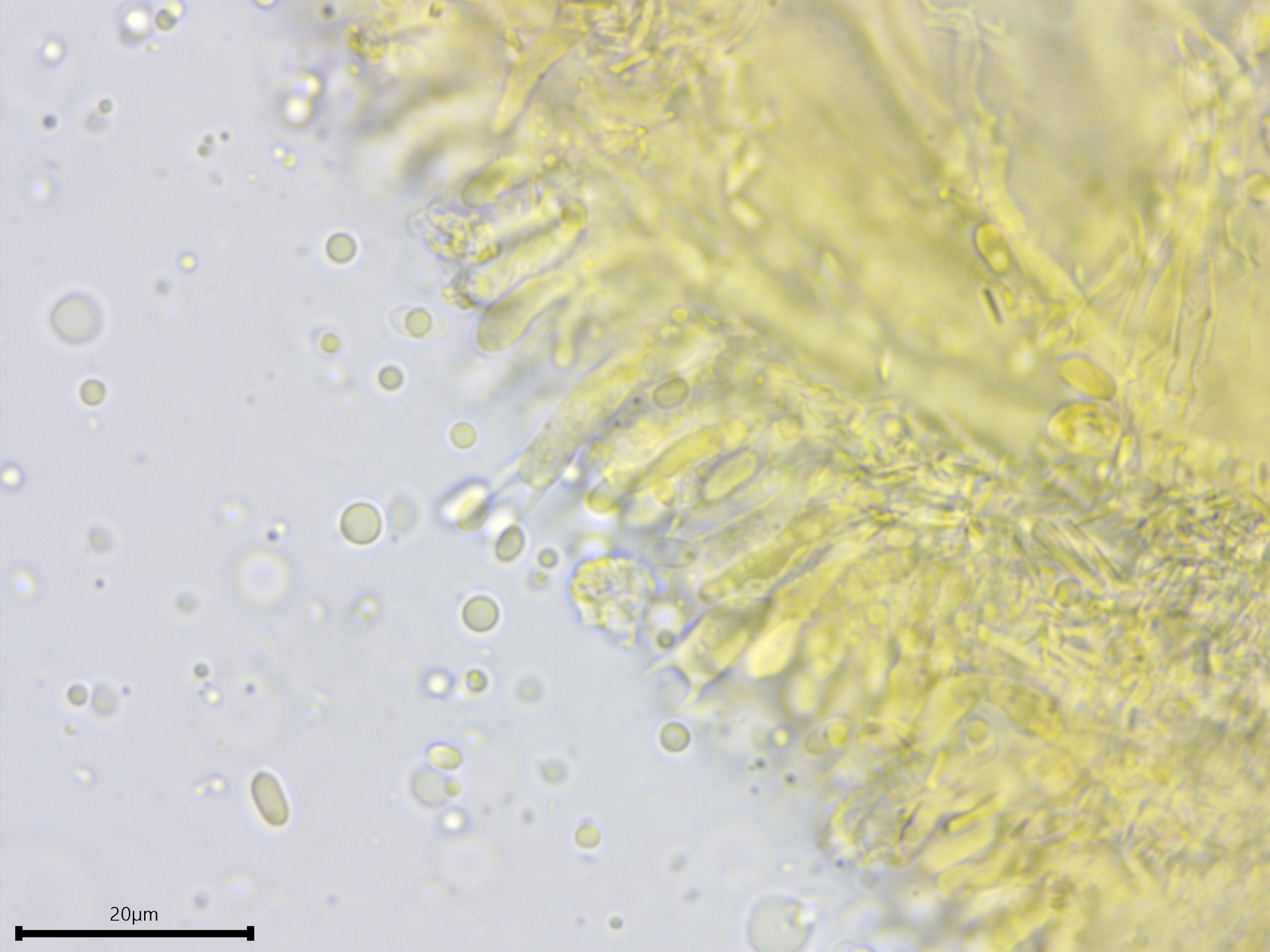
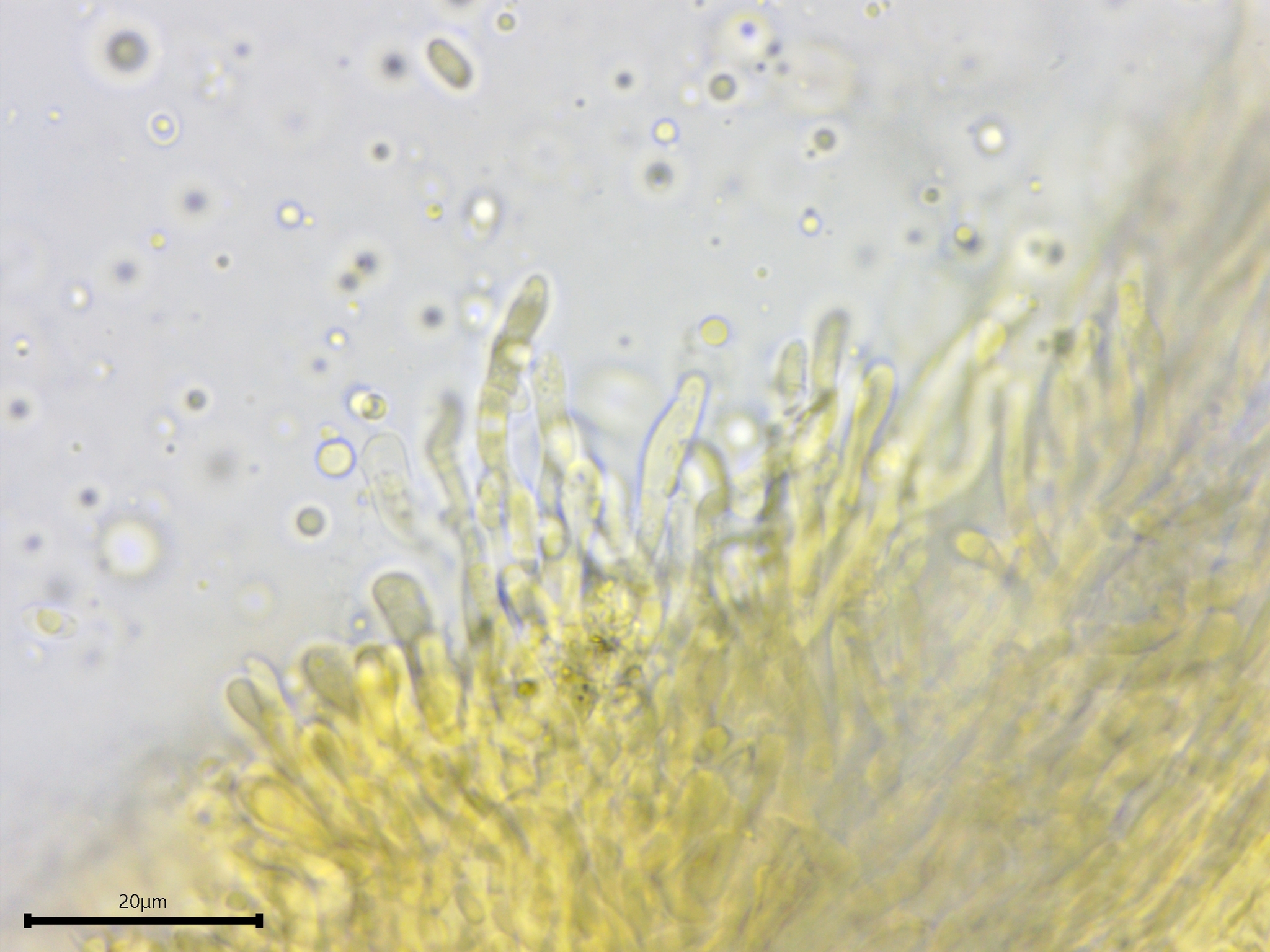
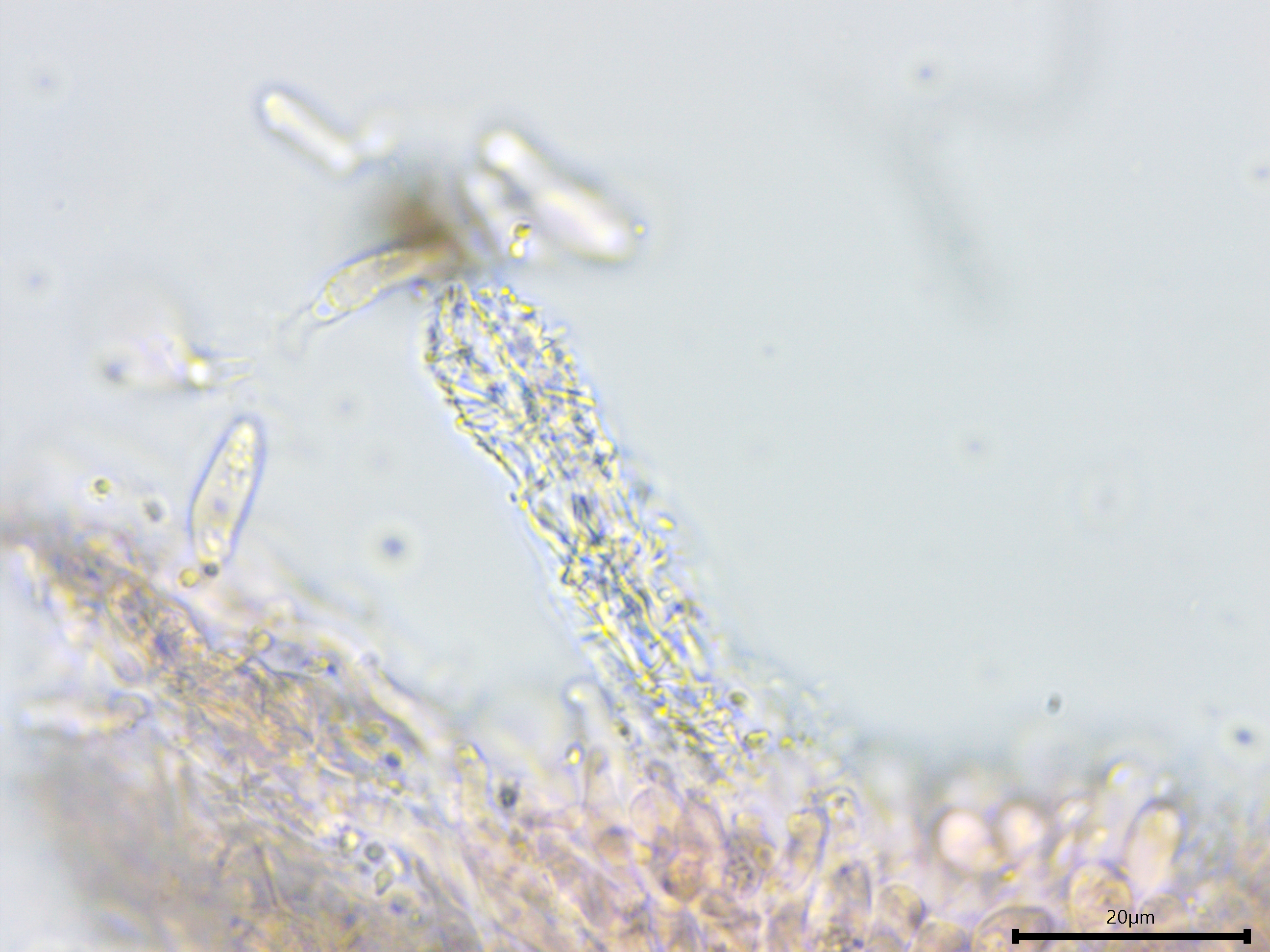
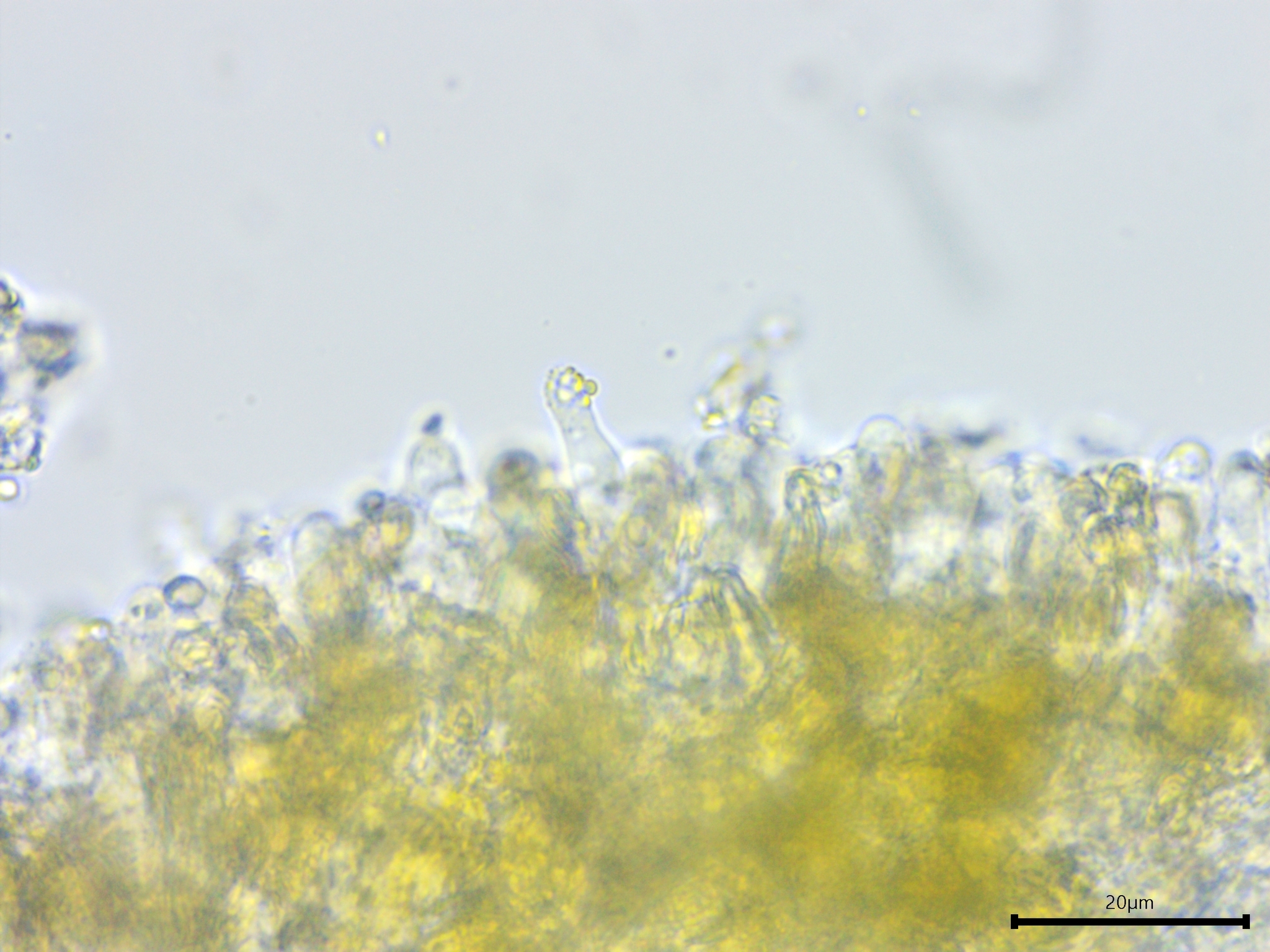
Sterile structures: Present as cystidioles projecting slightly beyond the hymenium, fusiform or slightly swollen at the apex, coated with a yellow resinous material that dissolves in KOH.
Sequences: ITS rDNA (OL755999), LSU rDNA (OL742442).
Notes: All measurements taken in 5% KOH stained with phloxine B. Pictures of this species with an extraordinarily bright yellow color abound on the internet. However, I have not come across any authoritative descriptions that portray it as such. For example, Nakasone described the color of Mycoacia uda as "pale yellow, light yellow, greyish yellow, light orange, greyish orange, brownish orange, yellowish brown, or brown" (1997). Mycoacia uda is sometimes reported as having spores that are 2.0–2.5 µm wide, but this is an error propagated from earlier studies whose text did not match the measurements in their drawings (Nakasone 1997).
Specimens Analyzed
ACD0388, iNat63490531; 23 October 2020; Eberwhite Nature Preserve, Washtenaw Co., MI, USA, 42.2737 -83.7683; leg. & det. Alden C. Dirks, ref. Bernicchia & Gorjón (2010); University of Michigan Fungarium MICH 352295.
References
Gorjón, S. P. (2020). Genera of corticioid fungi: Keys, nomenclature and taxonomy. Studies in Fungi, 5(1), 125–309.
Nakasone, K. K. (1997). Studies in Phlebia. Six species with teeth. Sydowia, 49(1), 49–79.
Links
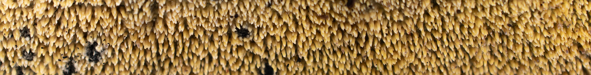
Pale yellow odontioid hymenium of Mycoacia uda.
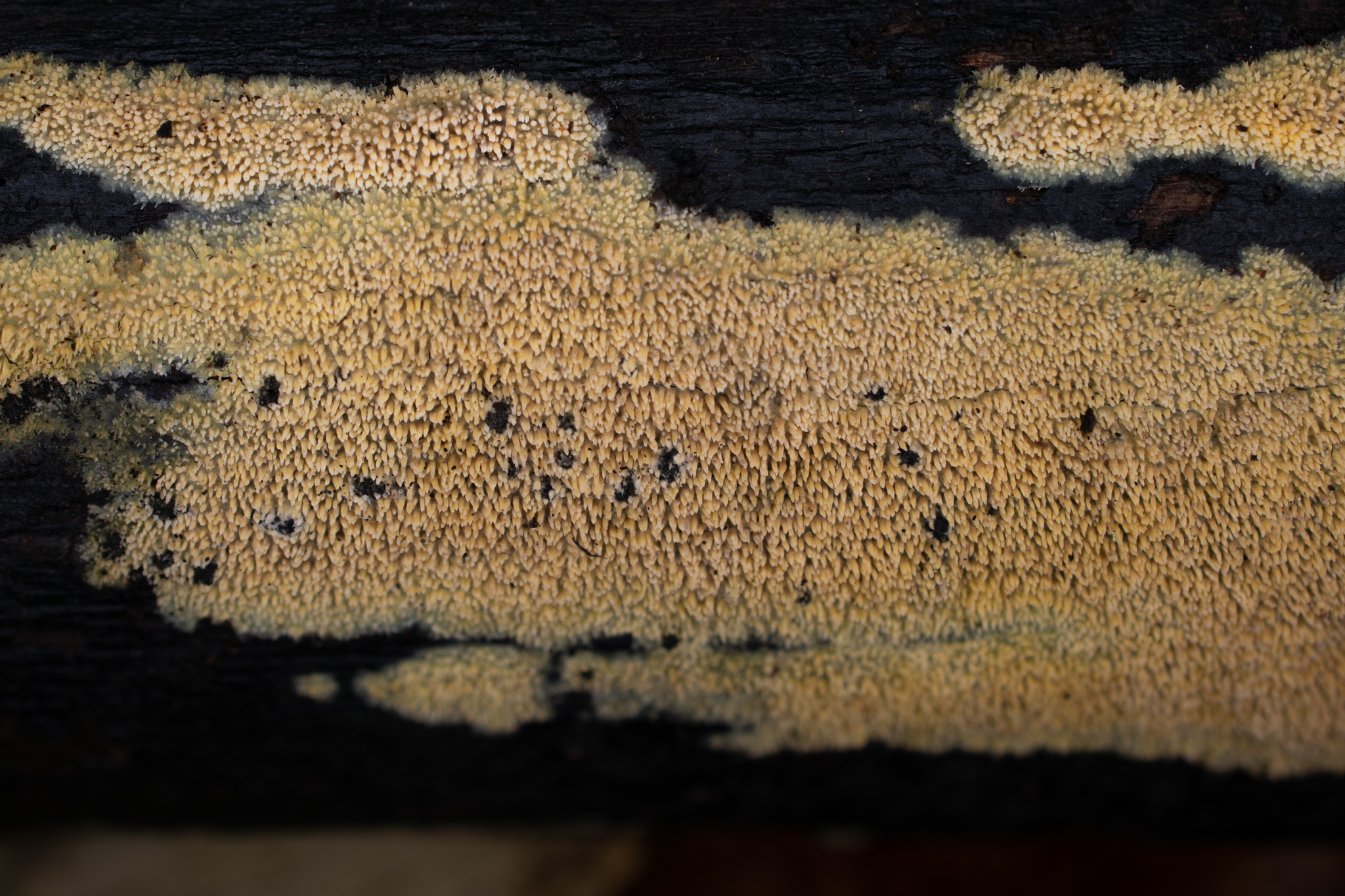
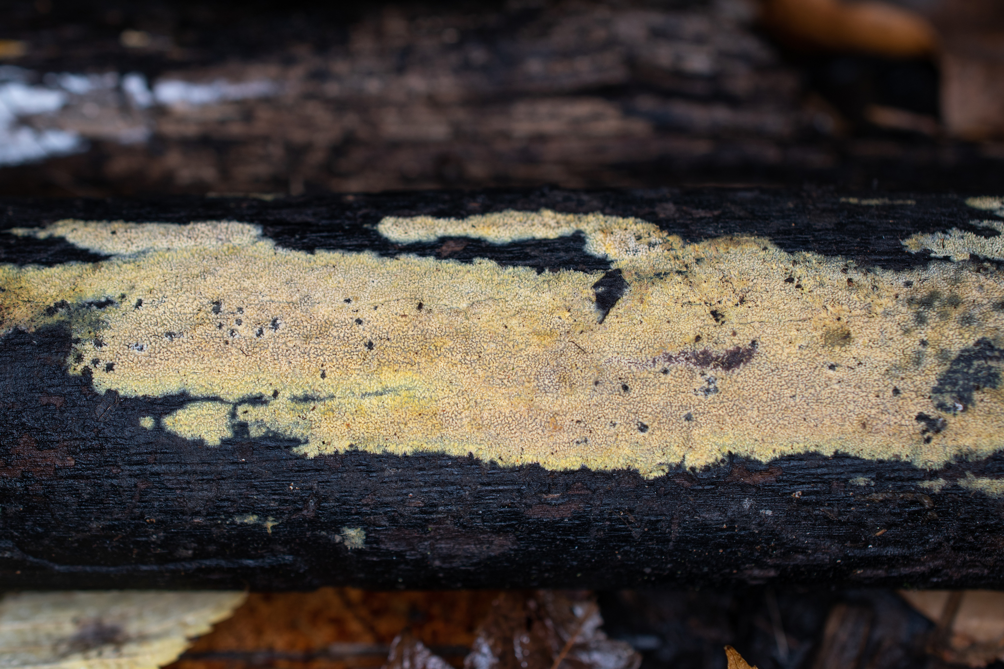
Notice the waxy consistency of the crust, especially evident at the margin.
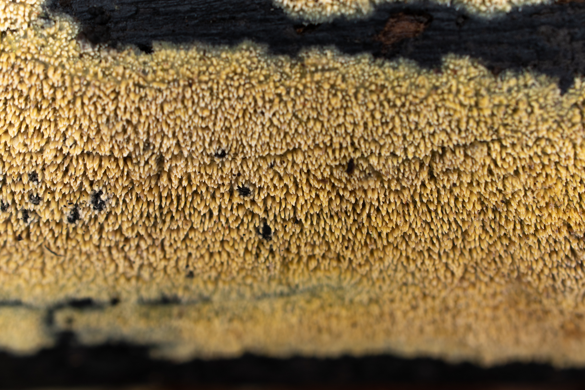
A closer look at the aculei with their paler tips.
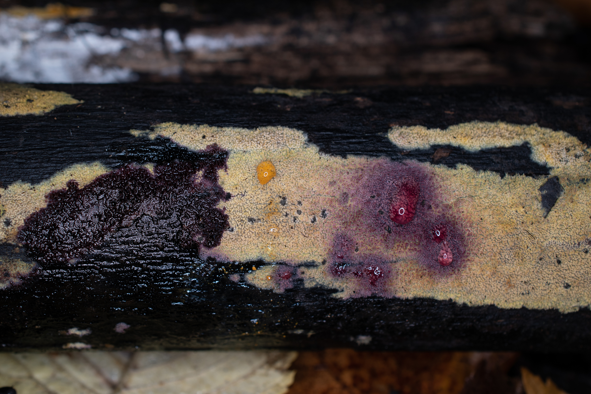
Ammonia and potassium hydroxide result in a distinctive purple color reaction.
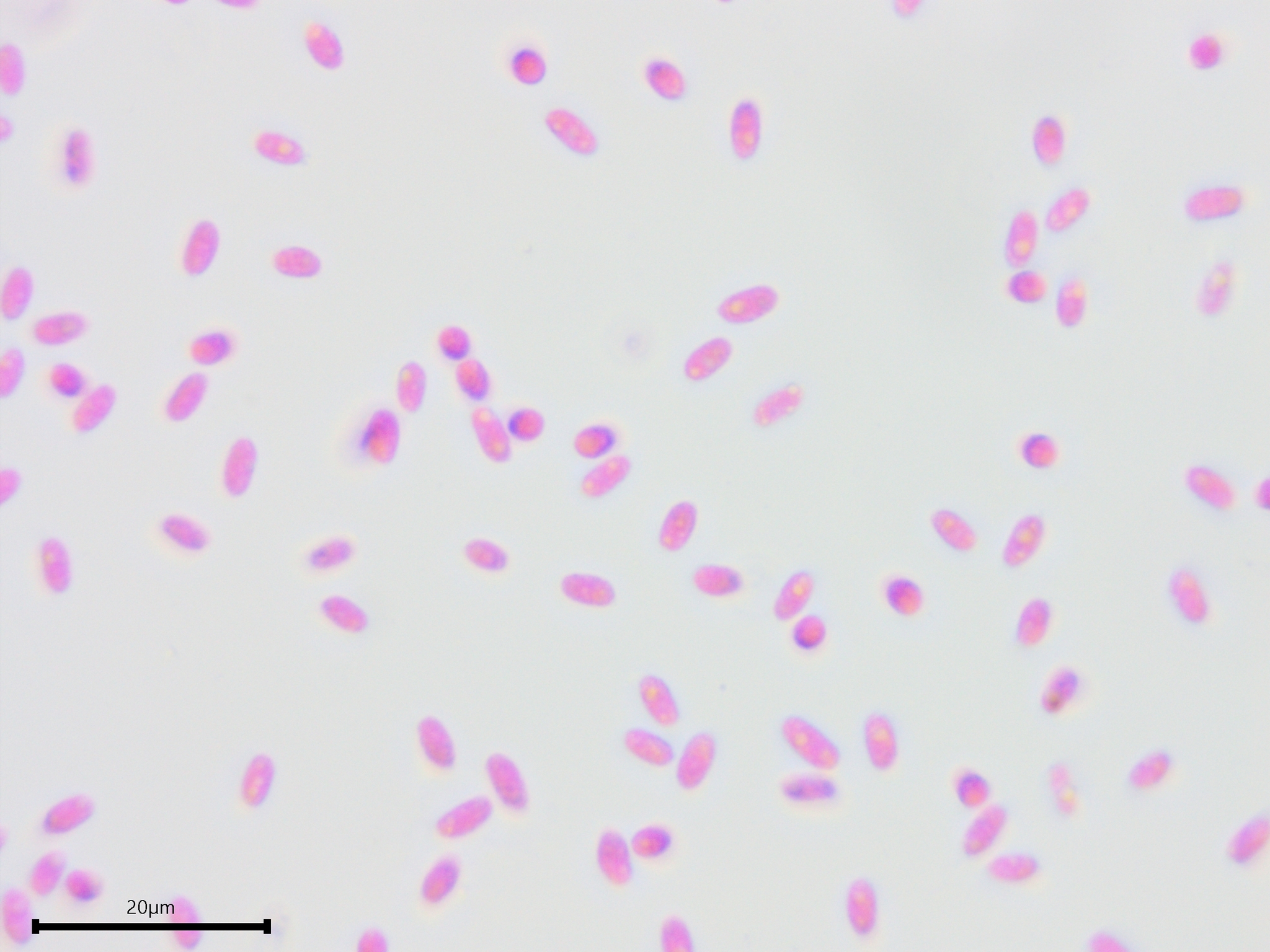
Cylindrical to narrowly cylindrical basidiospores.
Basidia.
Clamp connections.
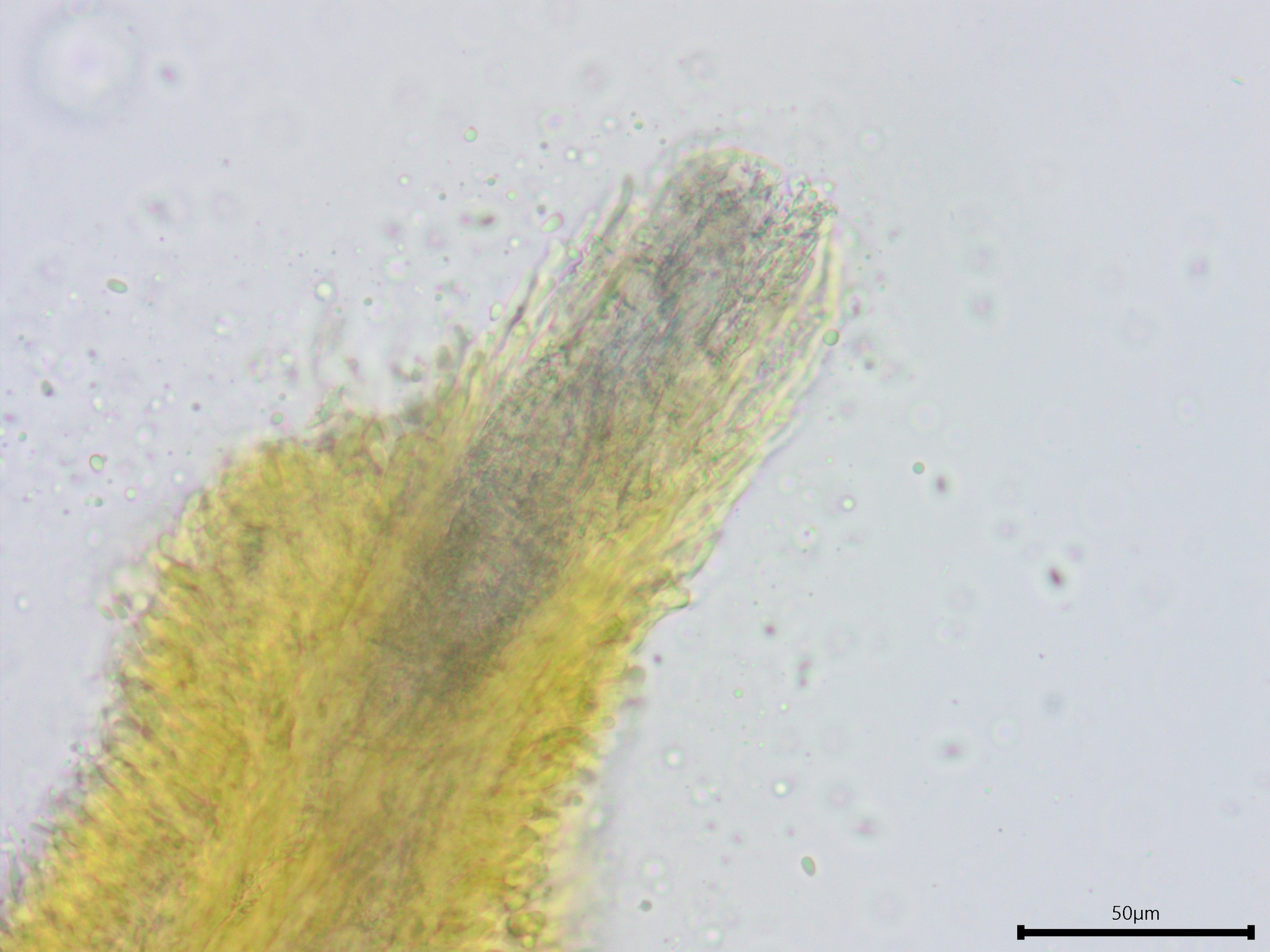
The hyphae in the trama of the aculei are encrusted with crystals and extend out of the tip as a blunt projection without hymenium.
A closer look at the acicular crytals on the tramal hyphae.
Yellow resinous cystidioles (visible in water).
Resinous capped cystidioles.