Subulicystidium brachysporum (P.H.B. Talbot & V.C. Green) Jülich
Basidiomycota: Agaricomycotina: Agaricomycetes: Trechisporales: Hydnodontaceae: Subulicystidium ![]()
![]()
Introduction
The genus Subulicystidium is characterized by amazing subulate (tapering to a point) cystidia with rectangular crystals that encircle the cystidia in rows or spirals. And poking out of the top of each cystidium is a hypha, which runs through the center of the barb-like structure. Who knows the adaptive function of these strange microscopic structures, but to me they look like they are for defensive purposes, perhaps against the many animals that graze on crusts.
The phylogeny below shows clusters of crusts labelled as Subulicystidium brachysporum spread throughout the tree, suggesting that S. brachysporum is a polyphyletic species complex in need of taxonomic revision.
Description
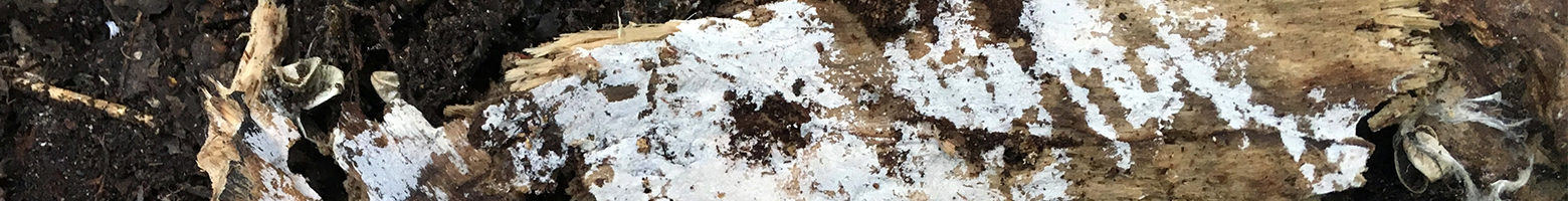
Ecology: Growing on the exposed wood of a fallen Salix (willow) in early summer, apparently with a global distribution.
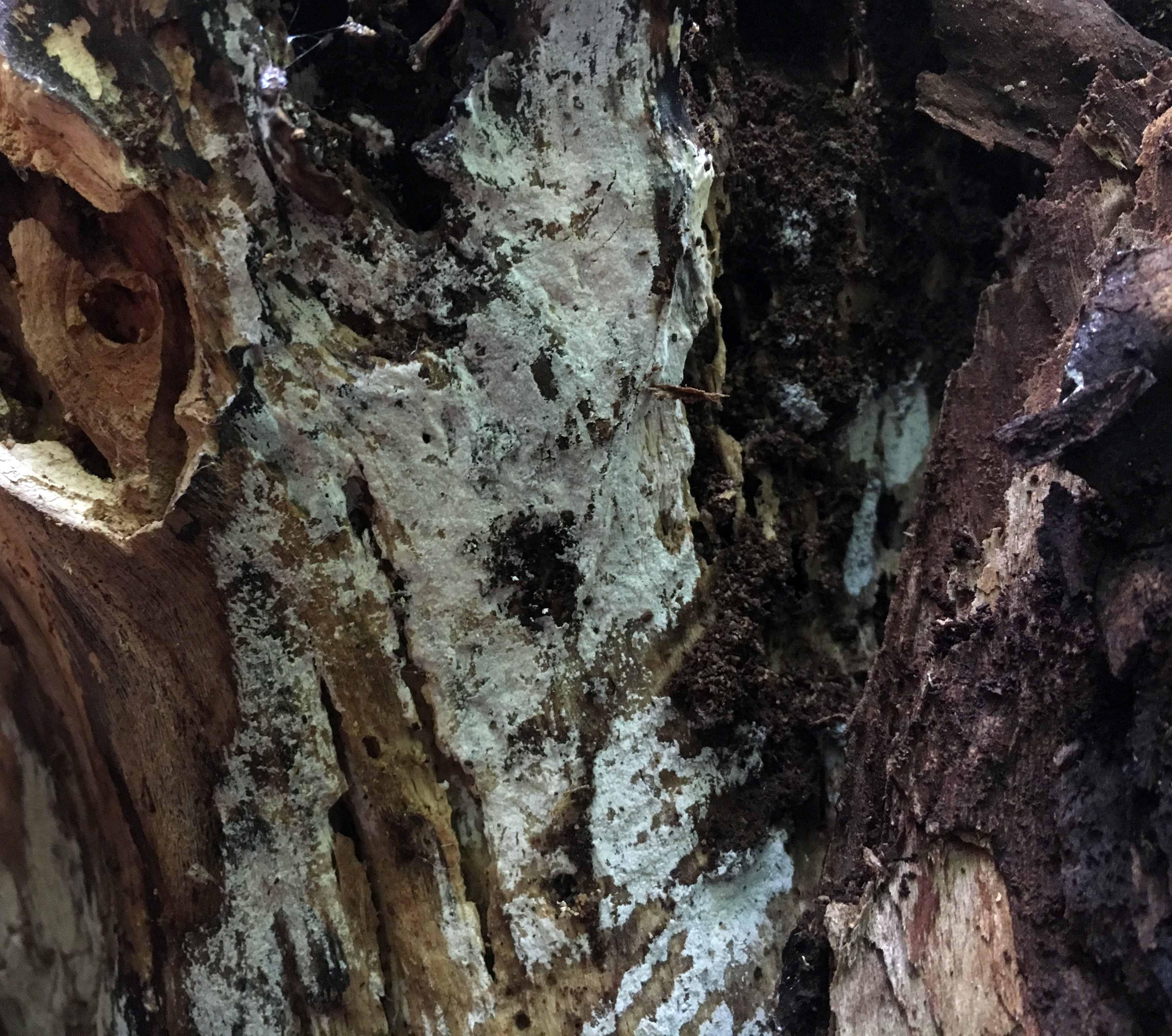
Basidiocarp: White, effused, resupinate basidioma with a smooth hymenophore.
Chemical reactions: NA
Spore print: NA
Hyphal system: Monomitic, encrusted, subicular hyphae somewhat thick-walled, clamped, width (3.6) 3.9–5.1 (5.3) µm, x̄ = 4.5 µm (n = 10 hyphae).
Basidia: Repetitive basidia with four sterigmata, about 15 ✕ 5 µm.
Basidiospores: Narrowly cylindrical to allantoid, thin-walled, hyaline, inamyloid; length (6.4) 7.5–8.8 (9.4) µm, width (2.2) 2.5–2.9 (3.2) µm, x̄ = 8.2 ✕ 2.7, Q (2.1) 2.7–3.3 (3.5); biguttulate (two equally sized oil droplets situated at the poles of the spore), rarely one or three guttules, diameter (1.2) 1.3-1.6 (1.6) µm (n = 30 basidiospores).
Sterile structures: Subulicystidia.
Sequences: ITS rDNA (MW448634).
Notes: All measurements taken in KOH stained with phloxine. Basidia were difficult to observe.
Specimens Analyzed
UWMA0061, MO324084; 23 June 2018; University of Wisconsin Arboretum, Dane Co., WI, USA, 43.0563 -89.4055; leg. & det. Alden Dirks, ref. Gorjón (2014), Ordynets et al. (2018); Kriebel Fungarium PULF24589.
References
Gorjón, S. P., Greslebin, A. G., & Rajchenberg, M. (2011). Subulicystidium curvisporum sp. nov. (Hymenochaetales, Basidiomycota) from the Patagonian Andes. Mycotaxon, 118, 47–52.
Gorjón, S. P. (2014). On-line key to Subulicystidium Parmasto. Online PDF (accessed December 12, 2020).
Ordynets, A., Scherf, D., Pansegrau, F., Denecke, J., Lysenko, L., Larsson, K.-H., & Langer, E. (2018). Short-spored Subulicystidium (Trechisporales, Basidiomycota): High morphological diversity and only partly clear species boundaries. MycoKeys, 35, 41–99.
Links
Subulicystidium brachysporum growing on the exposed, vertical wood of a fallen willow.
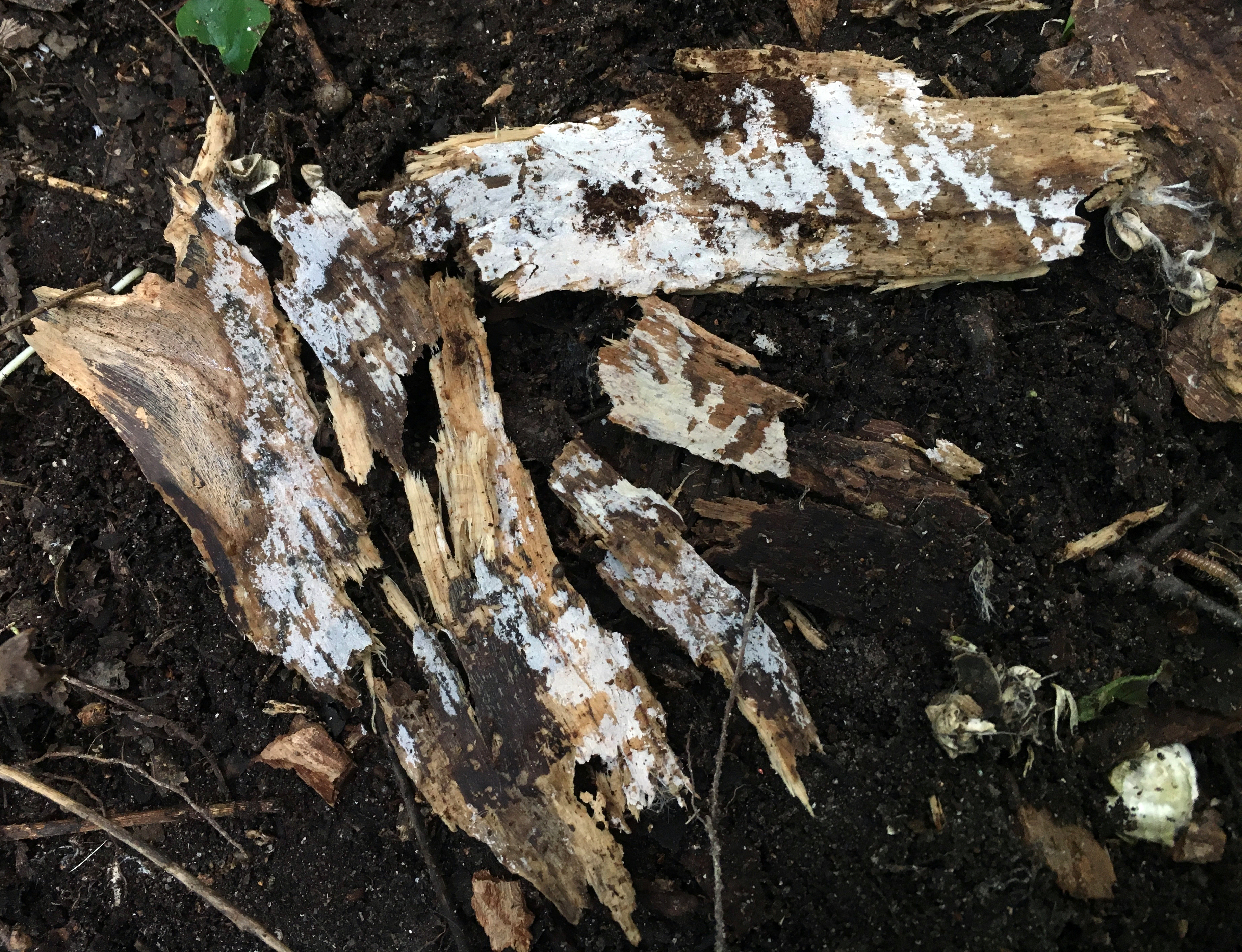
Sections of willow with the white, thin fruiting body of Subulicystidium brachysporum.
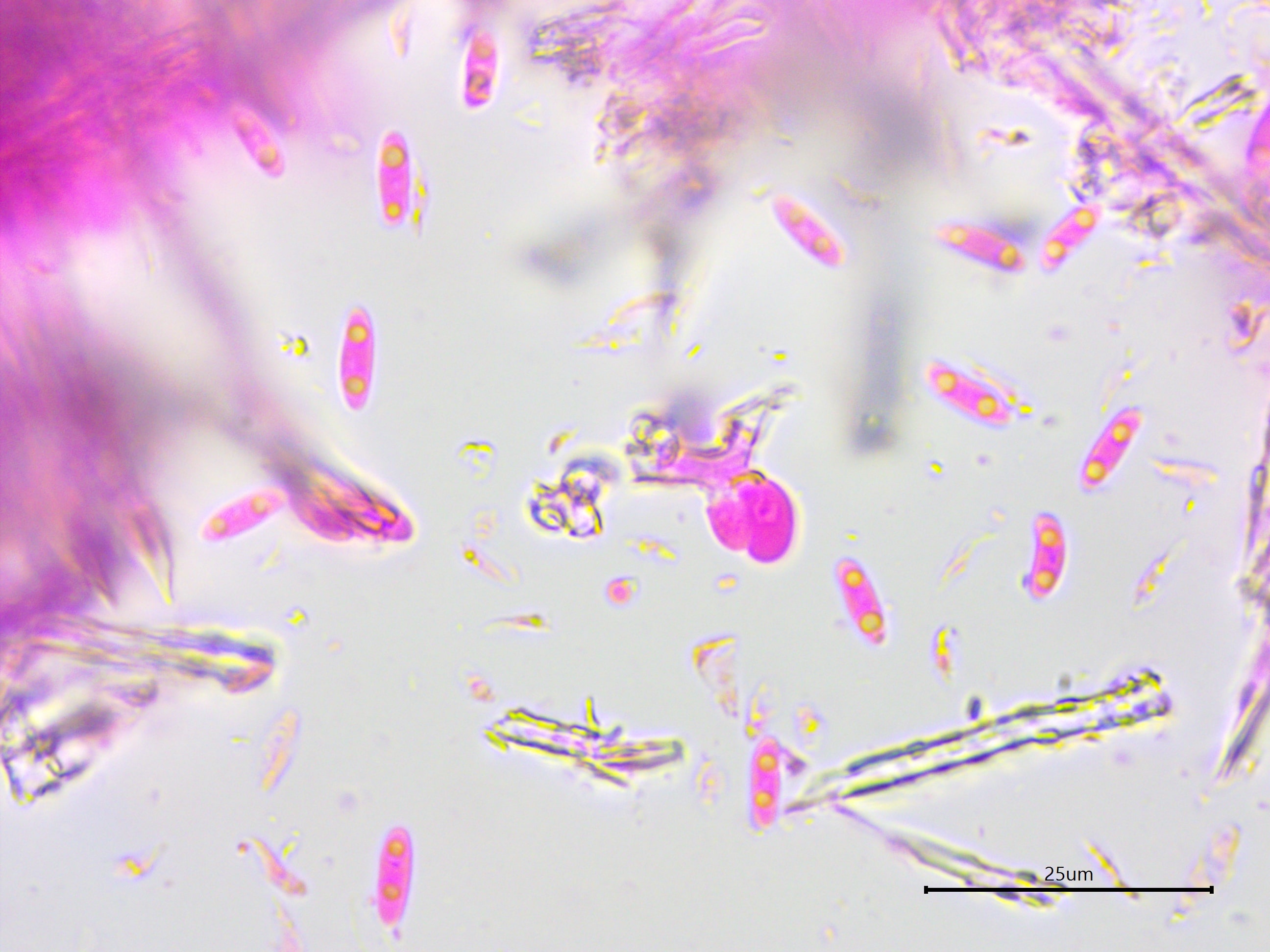
Biguttulate, narrowly cylindrical to allantoid spores.
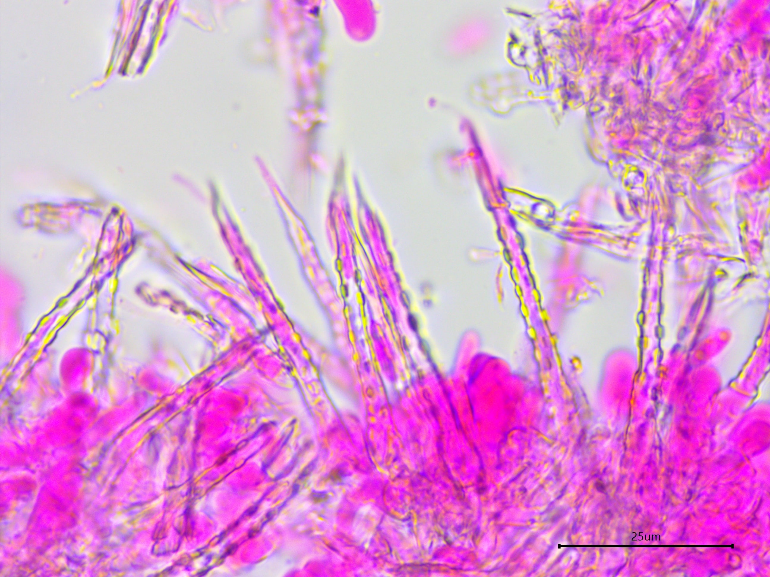
Needle-tipped subulicystidia.
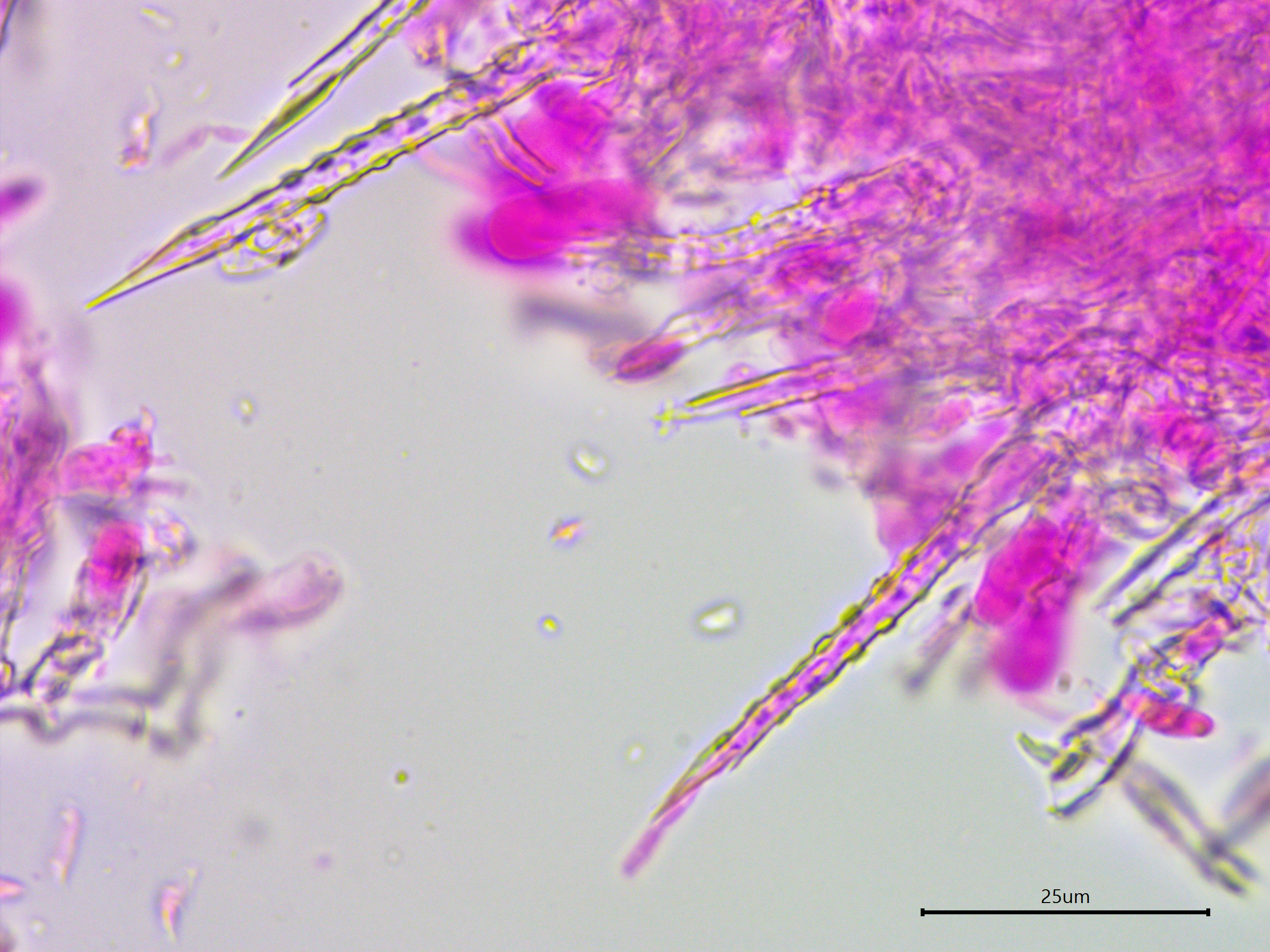
Here the hypha poking out of the subulicystidia can be seen more clearly.
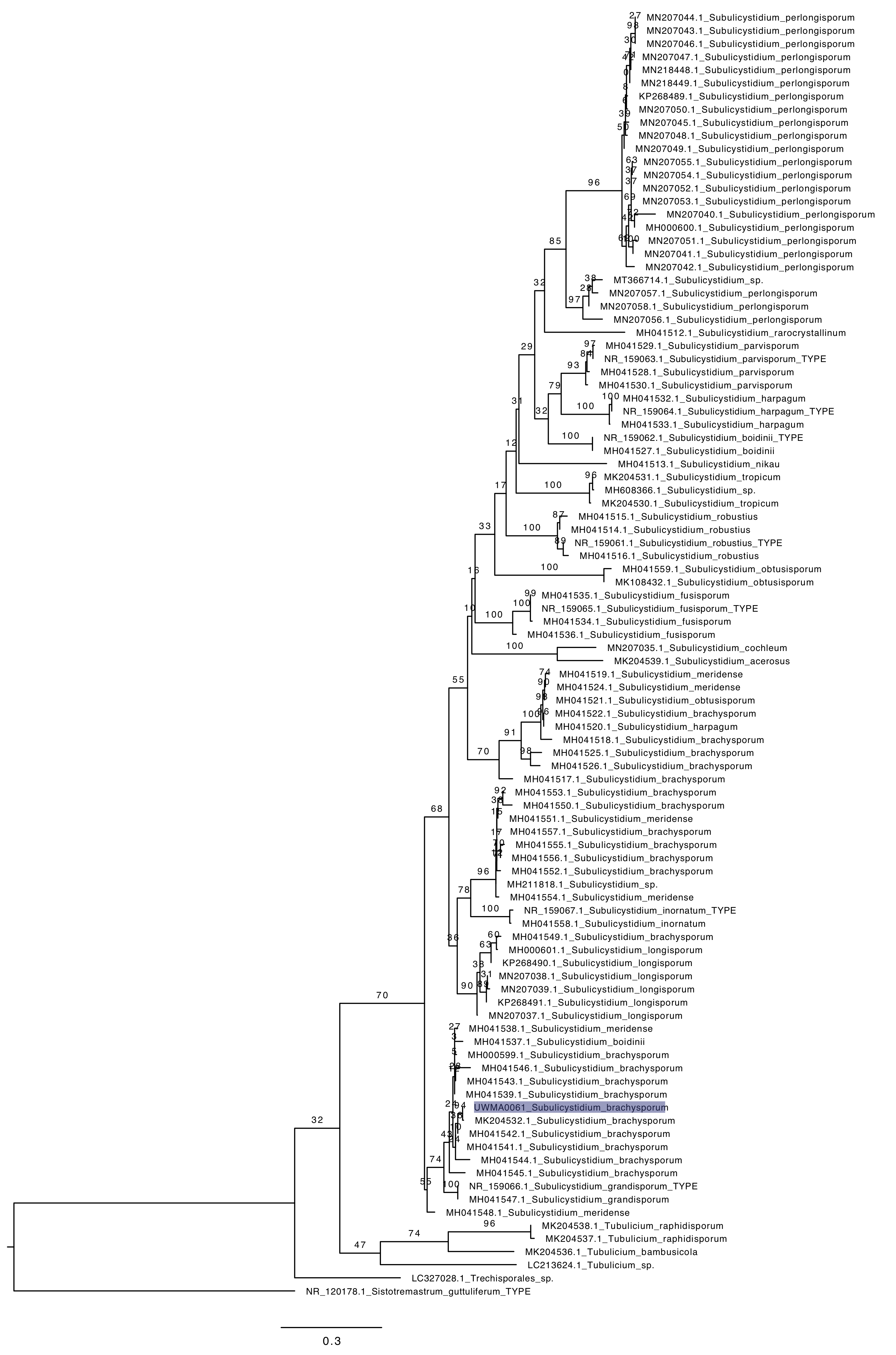
Phylogenetic tree of ITS rDNA sequences from the studied specimen (highlighted) and the top 100 similar vouchered specimens on GenBank. The sequences were processed with ITSx to remove the flanking SSU and LSU partial sequences, aligned in SeaView with MUSCLE, and made into a tree with RAxML using 100 bootsrap replicates and the GTRGAMMA substitution model. Sistotremastrum guttuliferum (type sequence) serves as the outgroup.