Basidiocarp

Bright yellow basidiome (Christian Schwarz, iNat15087060)

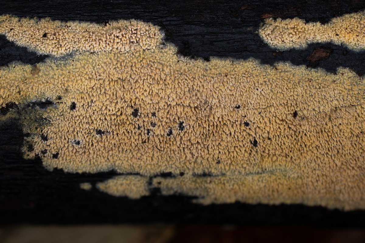
Basidiome (Jacob Kalichman, iNat112410038)

Basidiome (Dean Lyons, iNat329644821)

Close up of aculei (Damon Tighe, iNat184403152)

KOH and ammonia result in a purple reaction

Basidia

Cylindrical to narrowly cylindrical basidiospores.

Clamp connections

The hyphae in the trama of the aculei are encrusted with crystals and extend out of the tip as a blunt projection without hymenium

Acicular crytals on the tramal hyphae

Resinous capped cystidioles
Yellow Toothcrust
Sarcodontia uda (Fr.) Nikol.
Sarcodontia uda is a distinctive and beautiful yellowed toothed crust. Thanks to a pretty good number of DNA-barcoded observations on iNaturalist, it's evident that Sarcodontia uda is somewhat variable in color and configuration. The brightness and saturation of its yellow coloration varies from pale tan to neon, and the teeth can grow as an even stubble or clustered tufts.
With the basionym of Hydnum udum, this species has recently been passed around between the genera Phlebia, Mycoacia, Mycoaciella, and Sarcodontia. In molecular phylogenetic analyses, there is only weak statistical support for a relationship with Sarcodontia, so it may yet find itself in a new genus upon further study (Li et al. 2025).
Phlebioid crusts in the Meruliaceae family have received a lot of attention from the corticiologist Karen Nakasone. To give you a sense of the magnitude of the job (and hopefully a greater appreciation for the hard work of taxonomists), she analyzed almost 500 specimens in just one study on Sarcodontia uda and five other phlebioid crusts (Nakasone 1997)! And that's just one of many dozens of publications that she has published over her career. I'd be proud if I looked at that number of specimens in a lifetime.
Details
White-rot saprotroph on hardwood branches.
Odontioid to hydnoid; phlebioid (ceraceous when fresh, hardening upon drying); color yellowish, bright yellow to pale yellow or ochraceous, fading tannish brown when dry; aculei conical, up to 3 mm long, about 3 per mm, with sharp or frayed-looking (denticulate) tips, arising singularly or connected at the base with others from a thin, effused layer; margin abrupt or somewhat fimbriate, whitish.
Not determined.
Reacting purple to ammonia and potassium hydroxide, not absorbing iron salts (functionally negative).
White.
Temperate United States and Europe. View all sequenced specimens on iNaturalist.
Microscopy
Hyphal system: Monomitic; tramal hyphae hyaline, thick-walled, densely encrusted with fine, acicular crystalline material, 2–3 µm wide. Basidia: Cylindrical to clavate, terminal, reported as (16–)20–28(35) × 4–5 μm, tapering to 2–2.5 μm diam at base (Nakasone 1997), clamped at base, with four sterigmata. Basidiospores: Cylindrical to narrowly cylindrical, smooth, thin-walled, hyaline, inamyloid, sometimes guttulate; length (4.3) 4.8–5.5 (6.3) µm, width (2.2) 2.5–2.9 (2.9) µm, x̄ = 5.1 ✕ 2.7 µm, Q (1.6) 1.8-2.1 (2.3), x̄ = 1.9 (n = 30). Sterile structures: Present as cystidioles projecting slightly beyond the hymenium, fusiform or slightly swollen at the apex, coated with a yellow resinous material that dissolves in KOH (described as "resinous covered hyphal-end cells" by Nakasone [1997]); Nakasone (1997) reports "short, fusiform cystidia" that are "distinctive but often inconspicuous and difficult to observe".
Studied Specimens
ACD0388 (iNat63490531). 23 October 2020. Eberwhite Nature Preserve, Washtenaw Co., MI, USA, 42.2737 -83.7683. University of Michigan Fungarium (MICH352295). Sequences: OL755999 (ITS), OL742442 (LSU).
References
-
Li Y, Cao Y-F, Nakasone KK, Liu S-L, Huang M-R, He S-H. 2025. Species diversity, taxonomy, multi-gene phylogeny, and divergence times of Meruliaceae (Polyporales, Basidiomycota). Mycology 16:1180–1221. PDF Link
-
Nakasone KK. 1997. Studies in Phlebia. Six species with teeth. Sydowia 49:49–79. PDF Link
Citation
Dirks, Alden. 2026. Species profile for Sarcodontia uda (Fr.) Nikol. CrustFungi.Com. https://crustfungi.com/species/sarcodontia-uda/. Accessed 2026-04-04.