Ceraceous to gelatinous fresh basidiocarp

Dried basidiocarp, visible in bright light

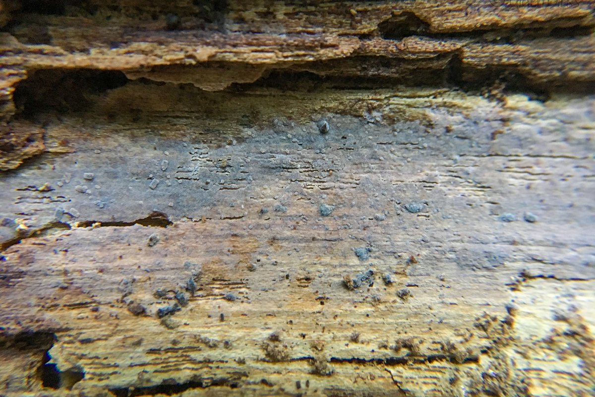
Gelatinous basidiocarp (Django Grootmyers, MO370363)

Basidium with two sterigmata

Developing basidium, weakly dextrinoid and ornamented basidiospores, and leptocystidium

Developing pleurobasidium

Ornamented basidiospores

Leptocystidium showing slight thickening of the cell wall towards the base
Xenasma praeteritum (H.S. Jacks.) Donk
The genus Xenasma consists of inconspicuous, whitish to bluish crusts. When fresh, they have a semitransparent, gelatinous appearance, but dry to a nearly invisible film. Unfortunately, these crusts can be as difficult to work with under the microscope as they are to see. The hyphae are small, gelatinized, and challenging to make out. The basidia and cystidia arise directly from this cryptic subiculum, which often appears embedded in the wood itself. While pleurobasidia are one of the defining features of Xenasma crusts, their hyphal origin is usually well hidden. But don't take my word for it — H. S. Jackson (1950) wrote the following about Xenasma:
The group as a whole is extremely difficult to study from dried material because of the gelatinous consistency when fresh and the vernicose ["brilliantly polished"] character when dry. Unless collections are dried quickly and were in active sporulating condition when collected, the basidia are difficult to make out. One is seldom able to view complete basidia or to trace them to the hyphae from which they originate.
Fortunately, there is one defining character of Xenasma crusts that is easy enough to see: ornamented spores. However, in all but one species, the spore ornamentation dissolves in KOH, further complicating identification. Xenasma praeteritum is the only one whose spores do not do this, a feature that makes it perhaps the easiest species to recognize in the genus.
Details
Presumably saprotrophic, on old, decorticated wood of deciduous trees.
Effused, smooth, ceraceous to gelatinous and somewhat transparent when fresh, drying to a subinvisible, pruinose film, margin undifferentiated and indeterminate, bluish.
Not determined, too insubstantial.
Not determined.
Not determined.
Reported from Eastern North America. View all sequenced specimens on iNaturalist.
Microscopy
Hyphal system: Monomitic, individual hyphae very difficult to discern, clamped. Basidia: Pleural (birooted), clavate to cylindrical, with two to four sterigmata (reported as three to five); length (23.8) 24–32.1 (34.9) µm, width (7.8) 8.0–9.4 (9.6) µm, x̄ = 28.1 ✕ 8.7 µm (n = 6 basidia). Basidiospores: Broadly ellipsoid, somewhat thick-walled, ornamented (asperulate) in Melzer's reagent and KOH, inamyloid or weakly dextrinoid; length (6.7) 7.0–8.0 (9.2) µm, width (5.0) 5.5–6.6 (7.4) µm, x̄ = 7.5 ✕ 6.1, Q (1.1) 1.2–1.3 (1.4) (n = 30 basidiospores). Sterile structures: Leptocystidia present, projecting well beyond the hymenium, thin-walled or somewhat thick-walled towards the base, at least 70 µm long and about 8 µm wide.
Studied Specimens
ACD0185 (iNat33478376). 24 September 2019. Escher Co-op Woods, Washtenaw Co., MI, USA, 42.2949 -83.7244. University of Michigan Fungarium (MICH352190). Sequences: OM009268 (ITS).
References
-
Jackson HS. 1950. Studies of Canadian Thelephoraceae: VI. The Peniophora rimicola group. Canadian Journal of Research 28:525–534. PDF Link
-
Liberta AE. 1960. A taxonomic analysis of section Athele of the genus Corticium. I. Genus Xenasma. Mycologia 52:884–914. PDF Link
Citation
Dirks, Alden. 2026. Species profile for Xenasma praeteritum (H.S. Jacks.) Donk. CrustFungi.Com. https://crustfungi.com/species/xenasma-praeteritum/. Accessed 2026-04-04.